


Article Figures & Data
Figures
- Fig. 1.
Analysis of recombinant UGT1A10 expression. (A) Western blot analysis of UGT1A10 protein in microsomes prepared from UGT1A10-6xHis-overexpressing stable CHO and HEK293 cell lines. Microsomes were boiled at 95°C for 10 minutes in the presence of 100 mM DTT prior to electrophoresis. (B) SDS-PAGE of microsomes under different denaturating conditions, followed by immunoblot analysis. DTT, electrophoresis after reduction with 100 mM DTT; Boiling, electrophoresis after boiling; No treatment, no DTT and boiling before electrophoresis.
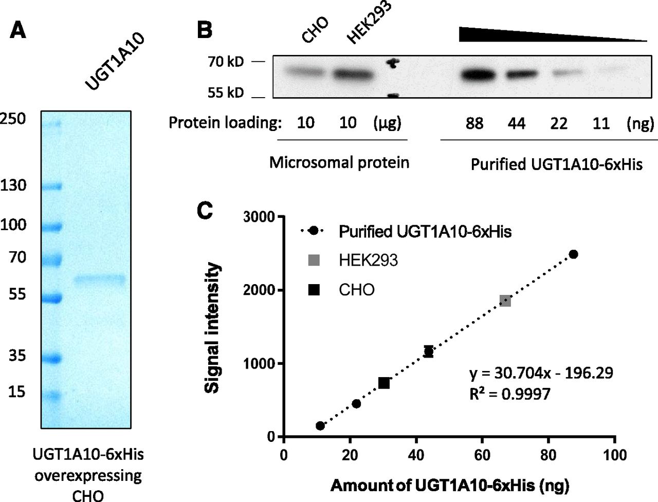
- Fig. 2.
Quantification of the levels of UGT1A10 in microsomes. (A) SDS-PAGE analysis of pure UGT1A10-6xHis extracted from microsome prepared from UGT1A10-6xHis-overexpressing stale CHO cells. The sizes of the molecular mass markers are indicated on the left in kilodaltons. (B) Chemiluminescent blot of dilution series of purified UGT1A10-6xHis and two unknown amounts of membrane-bound UGT1A10-6xHis. (C) The linear dynamic range of film-based detection for UGT1A10-6xHis. The graph shows a quantitative level of UGT1A10-6xHis protein for the corresponding chemiluminescence signal intensity. Each dot represents the average of duplicates and its values are expressed as the mean ± S.D.
- Fig. 3.
Kinetic analysis of the formation of M3G by UGT1A10. (A) HPLC analysis of M3G formation using microsomes prepared from UGT1A10-6xHis-overexpressing stable cell lines. Top, membrane-bound UGT1A10-6xHis expressed in CHO cells; middle, membrane-bound UGT1A10-6xHis expressed in HEK293 cells; bottom, membrane-bound UGT1A10-6xHis expressed in HEK293 cells with treatment with β-glucuronidase. (B) M3G formation by membrane-bound UGT1A10-6xHis enzymes in CHO cells (●) or in HEK293 cells (○), or by purified UGT1A10-6xHis protein (■) were determined at substrate concentrations of 0.1–20 mM. Kinetic parameters (kcat and KM) were determined by fitting the measured reaction rate data to the Michaelis-Menten kinetic equation using the Prism5.01 software. Each dot is the representative of the average of triplicates and its values are expressed as the mean ± S.D.
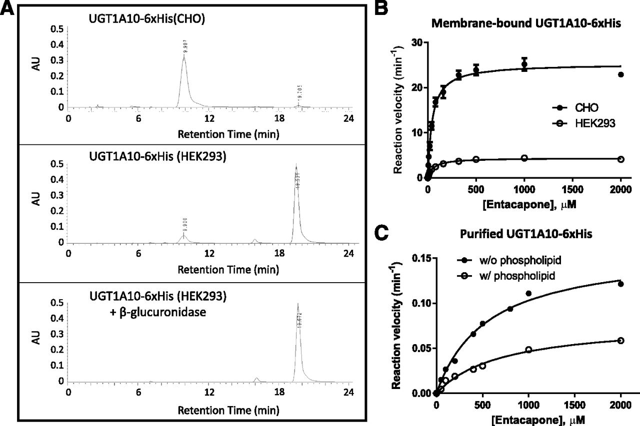
- Fig. 4.
Kinetic analysis of entacapone 3-O-glucuronide formation by UGT1A10. (A) HPLC analysis of the formation of entacapone 3-O-glucuronide using microsomes prepared from UGT1A10-6xHis-overexpressing stable cell lines. Top, membrane-bound UGT1A10-6xHis expressed in CHO cells; middle, membrane-bound UGT1A10-6xHis expressed in HEK293 cells; bottom, membrane-bound UGT1A10-6xHis expressed in HEK293 cells with treatment with β-glucuronidase. (B) Formation of entacapone 3-O-glucuronide by membrane-bound UGT1A10-6xHis protein in CHO cells (●), or in HEK293 cells (○) were determined at substrate concentrations of 3–2000 μM. (C) Formation of entacapone 3-O-glucuronide by purified UGT1A10-6xHis protein was determined at substrate concentrations of 3–2000 μM in the presence (●) or absence (○) of 1 mg/ml of phosphatidylcholine type X-E. Kinetic parameters (kcat and KM) were determined by fitting the generated reaction rate data to the Michaelis-Menten kinetic equation using the Prism5.01 software. Each dot refers to the average of duplicate or triplicates and its values are expressed as the mean ± S.D.


Tables
UGT1A10-6xHis KM kcat kcat/KM RCEa R2 μM min−1 min−1 · M−1 Membrane-bound_HEK293 56.9 ± 4.1 4.39 ± 0.07 7.71 × 10⁴ 1 0.987 Membrane-bound_CHO 45.7 ± 4.5 25.3 ± 0.6 5.53 × 10⁵ 7.17 0.992 Purified_CHO w/o phospholipid 703 ± 116 0.079 ± 0.005 1.12 × 10² 1.45 × 10⁻³ 0.991 Purified_CHO w/ phospholipid 554 ± 72 0.16 ± 0.01 2.88 × 10² 3.74 × 10⁻³ 0.995 ↵a Relative catalytic efficiency (kcat/KM).
Data Supplement
- Supplemental Data -
Supplemental Figure 1 - Immunoprecipitation assay for UGT1A10-6xHis
Supplemental Figure 2 - HPLC analysis of the formation of entacapone 3-O-glucuronide using microsomes prepared from UGT1A10-6xHis-overexpressing stable cell lines
Supplemental Figure 3 - The modeled van der Waals surface of the UGT1A10 protein structure, showing three solvent-accessible cysteine residues (C72, C183, and C277 in green color)
- Supplemental Data -


