


Abstract
Nicotine is the major addictive agent in tobacco; it is primarily metabolized by 5′-oxidation. 4-(Methylnitrosamine)-1-(3-pyridyl)-1-butanone (NNK), a potent lung carcinogen generated from nicotine during the curing of tobacco, is metabolically activated by P450 enzymes. P450 2A6 is the primary hepatic catalyst of nicotine metabolism and also catalyzes NNK α-hydroxylation, albeit less efficiently. It was previously reported that P450 2B6 catalyzed nicotine 5′-oxidation. The studies presented here investigate the relative importance of P450 2B6 as a catalyst of nicotine 5′-oxidation and NNK α-hydroxylation by human liver microsomes (HLMs). Radioflow high-performance liquid chromatography analysis and tritiated substrates were used to monitor the products of nicotine and NNK metabolism. The primary product of P450 2B6-catalyzed nicotine metabolism was the Δ1′(5′) iminium ion. The only other metabolite detected was nornicotine, the product of methyl oxidation, formed at about one-fourth the rate of the Δ1′ (5′) iminium ion. We determined that P450 2B6 was a much less efficient catalyst of nicotine 5′-oxidation than previously reported, with an estimated Km of 820 μM. In contrast, the Km of NNK α-hydroxylation was 33 μM. Experiments with P450 2A6- and P450 2B6-selective inhibitory antibodies did not support P450 2B6 as a significant catalyst of nicotine 5′-oxidation by HLMs, and it is unlikely that this enzyme contributes to nicotine metabolism in smokers who express P450 2A6. However, P450 2B6 contributed significantly to NNK metabolism in HLMs expressing both P450 2B6 and P450 2A6, suggesting a possible role for P450 2B6 in NNK metabolic activation.
Nicotine is the major addictive agent in tobacco (Benowitz, 1999), and NNK, a potent lung carcinogen, is generated from nicotine during the curing of tobacco. In tobacco users, nicotine is quickly metabolized to cotinine, a nonaddictive molecule. On the other hand, the carcinogenicity of NNK is dependent on its metabolism (Hecht, 1998). Nicotine and NNK are structurally similar compounds that are metabolized by a number of common enzymes (Hecht, 1998; Hukkanen et al., 2005; Jalas et al., 2005).
The primary pathway of nicotine metabolism in smokers is P450-catalyzed oxidation of the 5′-carbon (Hukkanen et al., 2005). The product of nicotine 5′-oxidation is 5′-hydroxynicotine, which exists in equilibrium with the nicotine Δ1′(5′) iminium ion (Fig. 1A). The Δ1′(5′) iminium ion is further metabolized to cotinine. This reaction may be catalyzed by cytosolic aldehyde oxidase (Brandänge and Lindblom, 1979) or, as we have recently reported, by P450 2A6 (Murphy et al., 2005; von Weymarn et al., 2005). P450 2A6 is the major catalyst of nicotine 5-oxidation in the human liver (Hukkanen et al., 2005). P450 2A6 also catalyzes two minor pathways of nicotine metabolism, oxidation of the N-methyl carbon and oxidation of the 2′-carbon (Fig. 1A) (Hecht et al., 2000; Murphy et al., 2005). Each of these pathways results in the formation of an iminium ion that is in equilibrium with the corresponding carbinolamine. The carbinolamine product of methyl oxidation is unstable and spontaneously generates nornicotine (Fig. 1A). At neutral pH, 2′-hydroxynicotine exists in about a 50:50 mixture with the amino ketone (Brandänge et al., 1983) (Fig. 1A).
It was previously reported that P450 2B6 catalyzes nicotine 5′-oxidation (Yamazaki et al., 1999). It is unknown whether P450 2B6 plays any role in the hepatic metabolism of nicotine. However, it might contribute to nicotine metabolism in the small number of individuals who do not express P450 2A6. The relative abundance of both 2A6 and 2B6 in the human liver is low. In many individuals P450 2B6 accounts for less than 1% of the total P450 present in the liver; the highest level reported was 4.1% (Hanna et al., 2000). P450 2A6 accounts for 1 to 10% of the total P450 in the liver (Pelkonen et al., 2000).
The metabolic activation of NNK occurs through P450-catalyzed α-hydroxylation at either the methyl carbon or methylene carbon position (Jalas et al., 2005) (Fig. 1B). Hydroxylation of the methyl carbon results in the formation of 4-(3-pyridyl)-4-oxo-butyl(diazohydroxide) and formaldehyde. The diazohydroxide can react with water to form either 4-hydroxy-4-(3-pyridyl)butanol (HPB) or pyridyl-oxobutylate DNA to form DNA adducts. Methylene carbon α-hydroxylation generates methane diazohydroxide and 4-oxo-4-(3-pyridyl)butanal (OPB), which can be further oxidized to the acid (OPBA). Methane diazohydroxide can either methylate DNA to form adducts or react with water to form methanol.
Several hepatic P450s have been investigated as catalysts of NNK α-hydroxylation. The relative importance of each of these was recently reviewed (Jalas et al., 2005). Specifically, chemical inhibitors, inhibitory antibodies, and correlations with P450-specific substrates have been used to assess the catalytic contributions of 2A6, 3A4, 1A2, 2D6, 2E1, and 2C8 in human liver microsomes (HLMs). P450s 2A6 and 1A2 have been suggested to be the enzymes involved in human hepatic NNK metabolism (Jalas et al., 2005). However, both of these enzymes are relatively poor catalysts with Km values greater than 300 μM. The role of P450 2B6 in the catalysis of NNK α-hydroxylation has not been investigated. The goal of this study was to investigate the relative importance of P450 2B6 as a catalyst of nicotine 5′-oxidation and NNK α-hydroxylation.
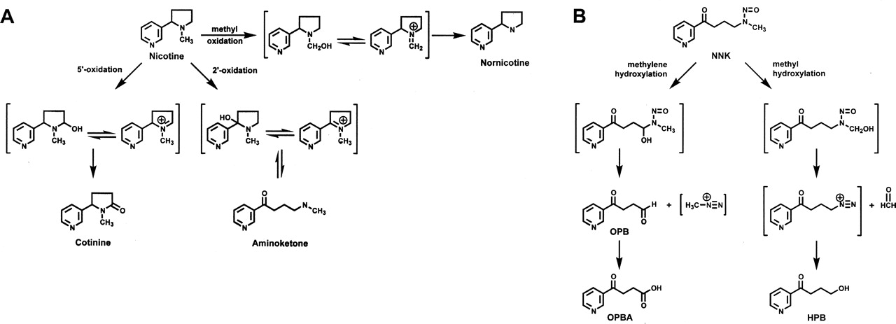
Nicotine oxidation (A) and NNK α-hydroxylation pathways (B).
Materials and Methods
Chemicals, Reagents, and Enzymes.S-Mephenytoin was purchased from Toronto Research Chemicals (North York, ON, Canada) and 5-phenyl-5-ethylhydantoin (Nirvanol) was purchased from Ultrafine Chemicals (Manchester, UK). [5-3H]NNK (11 Ci/nmol, 99.9% radiochemical purity) was purchased from Moravek Biochemicals (Brea, CA). (S)-[5-3H]Nicotine was prepared and purified as previously described (Murphy et al., 2005). All other chemicals were purchased from Sigma-Aldrich (St. Louis, MO). P450 2B6 and P450 2A6 Supersomes and P450 2B6 and P450 2A6 antibodies were purchased from BD Biosciences Discovery Labware (Bedford, MA).
P450 2A6 and P450 2B6 Content of HLMs. Liver samples 1 and 2 were a gift from Dr. Rory Remmel (University of Minnesota, Minneapolis, MN). All other liver tissue samples were provided by Dr. F. Peter Guengerich (Vanderbilt University, Nashville, TN). The preparation of the HLMs was as previously described (Fowler et al., 1994). HLM HH2 was obtained from BD Biosciences Discovery Labware. The relative expression of P450 2A6 and P450 2B6 in each HLM sample was determined by Western blot analysis. HLM samples (60 μg for P450 2B6 blot, 1–2 μg for P450 2A6 blot) were subject to polyacrylamide gel electrophoresis using 10% polyacrylamide gels (Guengerich, 1994). Standard amounts (0.022–0.5 pmol) of partially purified Escherichia coli-expressed P450 2B6 (a gift from Dr. Paul Hollenberg) or P450 2A6 (von Weymarn et al., 2005) were applied to each gel to generate a standard curve. Proteins were transferred to a polyvinylidene difluoride membrane overnight at 20 V and 4°C. Following transfer, the protein gel was stained with Gel Code Blue (Pierce Chemical, Rockford, IL) to confirm even transfer. The polyvinylidene difluoride membrane was blocked for 1 h in 3% nonfat milk and then incubated 1 h with either P450 2B6 or P40 2A6 antibody diluted 1:1000 in PBST. After three rinses with PBST for 10 min each, the membrane was incubated with secondary antibody diluted 1:1000 for 1 h (horseradish peroxidase-conjugated anti-rabbit IgG antibody for P450 2B6 and horseradish peroxidase-conjugated anti-mouse IgG for P450 2A6). After three 10-min rinses with PBST, the immunoreactive bands were visualized by a 1-min treatment with ECL Plus (GE Healthcare, Little Chalfont, Buckinghamshire, UK). Quantification of the bands was by densitometry using a STORM 840 (GE Healthcare) scanning densitometer and ImageQuant software. Two independent analyses were carried out for each HLM sample to determine P450 2B6 and P450 2A6 levels.
Coumarin 7-Hydroxylase and (S)-Mephenytoin N-Demethylase Activity. HLM samples were analyzed for coumarin 7-hydroxylase activity as a measure of P450 2A6 activity and (S)-mephenytoin N-demethylation as a measure of P450 2B6 activity. Coumarin 7-hydroxylase activity was determined as previously described (von Weymarn et al., 1999). The analysis of (S)-mephenytoin N-demethylation was a modification of previously described methods (Heyn et al., 1996; Ko et al., 1998). HLM samples (0.3 mg) were incubated in 0.1 M sodium phosphate buffer, pH 7.4, with an NADPH-generating system (0.4 mM NADP+, 100 mM glucose 6-phosphate, and 0.4 units ml–1 glucose-6-phosphate dehydrogenase), and 100 μM S-mephenytoin in a final volume of 0.5 ml for 45 min at 37°C. Duplicate reactions were run for each HLM sample, with and without 25 μM sulfaphenazole, an inhibitor of P450 2C9 activity (Ko et al., 1998). Reactions were terminated by the addition of 100 μl of ice-cold acetonitrile. Then, the internal standard (415 pmol of phenytoin) was added and the mixture was extracted twice with 0.5 ml of methylene chloride. The combined organic fractions were evaporated to dryness under a stream of N2 (gas). The residue was dissolved in 200 μl of the HPLC mobile phase [69:24:7 ratio of 20 mM sodium perchlorate (pH 2.5)/methanol/acetonitrile] and injected onto an Alltech Nucleosil C18 column (5 μm, 150 mm × 4.6 mm; Alltech Associates, Deerfield, IL). Mephenytoin and its metabolites were eluted isocratically with a flow rate of 1 ml/min. Detection was by UV absorbance at 204 nm with a Shimadzu SPD-10ADvp spectrophotometer (Shimadzu, Kyoto, Japan). Nirvanol, mephenytoin, and phenytoin eluted at 14.5, 26.9, and 55.6 min.
NNK α-Hydroxylation. The NNK α-hydroxylation activity of each HLM sample was determined by radioflow HPLC as previously described (Wong et al., 2005), except that no sodium bisulfite was included in the reaction and a different HPLC column was used. Reactions were run in the absence or presence of 2 pmol of inhibitory P450 2A6 or 2B6 antibodies for 30 min at 37°C. The HPLC column used was a Phenomenex (Torrance, CA) Gemini C18 column (5 μm, 250 × 4.60 mm) eluted with a gradient from 100% A [20 mM sodium phosphate (pH 7) containing 1 mM sodium bisulfite] to 70% A over 30 min, and then to 50% A in 10 min; B was 95% methanol. The eluant flow rate was 0.7 ml/min; the scintillant [Picofluor 40, PerkinElmer Life and Analytical Sciences, Boston, MA) flow rate was 3.0 ml/min.
(S)-Nicotine 5-Oxidation. The products of nicotine metabolism by HLM samples were analyzed by radioflow HPLC as previously described (Murphy et al., 2005). Each HLM sample (0.3 mg) was incubated with (S)-[5-3H]nicotine (100 μM, 0.1 μCi nmol–1) and an NADPH-generating system in 50 mM Tris, pH 7.4 for 30 min with or without P450 2A6 antibody (2 pmol). To determine the kinetic parameters of nicotine 5′-oxidation, the concentrations of nicotine used were 10, 20, 50, 100, 500, or 1000 μM (the specific activity was from 1.0 to 0.01 μCi/nmol). Each sample was analyzed in duplicate. P450 2B6 Supersomes (5–20 pmol) were incubated with either [5-3H]NNK (2.5–150 μM) or (S)-[5-3H]nicotine (50–1000 μM) for 30 min at 37°C. The products of NNK α-hydroxylation were analyzed by radioflow HPLC as described above for HLM samples. Nicotine 5′-oxidation was analyzed both in the absence and presence of cytosol, as a source of aldehyde oxidase, and the kinetic parameters were determined as previously described (Murphy et al., 2005).
Results and Discussion
P450 2B6 was previously reported to be a catalyst of nicotine 5′-oxidation, measured as cotinine formation in the presence of cytosol (Yamazaki et al., 1999). The Km and Vmax values reported were 105 μM and 8.2 nmol/min/nmol P450. In the present study, using tritium-labeled nicotine, we monitored the formation of both the nicotine Δ1′(5′) iminium ion and cotinine in the presence of cytosol. Sufficient cytosol was added to convert all of the Δ1′(5′) iminium ion to cotinine. Cotinine was not detected in the absence of cytosol. Nornicotine was also detected as a product of P450 2B6-catalyzed nicotine metabolism, accounting for 20% of the total metabolites formed. The estimated apparent Km for P450 2B6-catalyzed nicotine 5′-oxidation was determined to be 820 ± 190 μM and Vmax was 24 ± 4 nmol/min/nmol. However, the enzyme was not saturated at 1 mM nicotine, the highest concentration used. The large difference between the kinetic values reported here and those reported by Yamazaki et al. (1999) may be due to the addition of insufficient cytosol in the earlier experiments.
Any role for P450 2B6 as a catalyst of nicotine 5′-oxidation in human liver was investigated by determining the relative effect of P450 2A6- and P450 2B6-selective antibodies on this reaction in HLM samples. The samples analyzed were selected due to their relatively high level of P450 2B6 protein. The concentration of P450 2B6 in five samples was 3 to 4.4 pmol/mg and accounted for as much as 4% of the total P450 content (Table 1). Despite these relatively high P450 2B6 concentrations, the amount of P450 2A6 protein present in these samples was still 4- to 45-fold greater (Table 1). In these samples, P450 2A6 protein content and P450 2A6 activity, measured as the rate of coumarin 7-hydroxylation, were positively correlated (r = 0.70). With the exception of HLM sample 39, P450 2B6 content was positively correlated (r = 0.68) with the P450 2B6-catalyzed reaction S-mephenytoin N-demethylase. P450 2B6 activity in HLM sample 39 may have been lost during the preparation of microsomes.
P450 2B6 and 2A6 content relative to the rate of nicotine 5′-oxidation by human liver microsomes
The products of nicotine metabolism generated by nine HLM samples were determined by radioflow HPLC analysis. A typical trace is presented in Fig. 2. The major radioactive metabolite coeluted with the nicotine Δ1′(5′) iminium ion and was quantitatively converted to cotinine upon the addition of cytosol, a source of aldehyde oxidase. Interestingly, a significant amount of cotinine was also formed in the absence of cytosol. Cotinine was a detectable metabolite in seven of the nine HLM samples, accounting for as much as 39% of the nicotine metabolites. These data support a role for P450 enzymes as catalysts of the conversion of the Δ1′(5′) iminium ion to cotinine; this role was previously suggested by Shigenaga et al. (1988), and we have reported that both P450 2A6 and P450 2A13 catalyze this reaction (von Weymarn et al., 2005). No nicotine metabolites other than cotinine and the Δ1′(5′) iminium ion were detected in any samples. The rate of nicotine 5′-oxidation ranged from 1.5 to 223 pmol/mg/min. The lowest rate of nicotine metabolism was in HLM sample HH2, which contains no P450 2A6. The kinetic parameters of nicotine 5′-oxidation were determined for four HLM samples (2, 130, 131, and 141). The Km values obtained, 177 ± 11, 170 ± 50, 132 ± 19, and 110 ± 16, were quite similar to the value of 144 μM we reported recently for P450 2A6-catalyzed nicotine 5′-oxidation (Murphy et al., 2005).
At a nicotine concentration of 100 μM, P450 2A6-selective antibody inhibited nicotine 5′-oxidation significantly (83–97%) in all samples with P450 2A6 activity (Table 1). The effect of inhibitor antibodies on the rate of 5′-oxidation of 500 μM nicotine was determined for six HLM samples (1, 2, 130, 131, 134, and 141). Inhibition by P450 2A6 antibody ranged from 64 to 87% and inhibition by P450 2B6 antibody was from 0 to 35%. As one might predict, the samples that were inhibited the least by P450 2A6-selective antibody were inhibited the most by P450 2B6-selective antibody. Therefore, total inhibition by these two antibodies ranged from 80 to 99%. These data are consistent with P450 2A6 and P450 2B6 as the only enzymes that catalyze a significant level of nicotine metabolism in the liver. However, due to the much lower abundance of P450 2B6 and its rather high Km for nicotine 5′-oxidation relative to P450 2A6, it seems unlikely that P450 2B6 would play a significant role in nicotine metabolism in vivo, except in individuals who do not express P450 2A6.

Radioflow HPLC analysis of nicotine metabolism by human liver microsomes.
P450 2A6 is a catalyst of NNK metabolism; however, it is not a particularly efficient one. The rat P450 2B1 catalyzes NNK α-hydroxylation (Jalas et al., 2005). Therefore, it was of interest to determine the catalytic efficiency of the human ortholog, P450 2B6, for NNK α-hydroxylation. The major product of P450 2B6-catalyzed NNK α-hydroxylation was HPB, the product of methyl hydroxylation (Fig. 1B). HPB formation accounted for 85% of NNK α-hydroxylation by P450 2B6; OPB formation accounted for the remaining 15%. The kinetic parameters for total NNK α-hydroxylation were 33 ± 0.7 μM and 0.18 ± 0.01 pmol/min/pmol for the Km and Vmax. The Km was about 10 times lower and Vmax/Km 5 to 10 times higher than the values reported for P450 2A6 (Jalas et al., 2005).
The rates of NNK methyl and methylene hydroxylation were determined for 10 HLM samples (Table 2). The concentration of NNK used, 80 μM, is more than twice the Km of P450 2B6 but is still much less than the Km of 2A6 and was chosen to detect a possible contribution of P450 2B6 to NNK α-hydroxylation by HLM. The rates of NNK α-hydroxylation varied 5-fold among the HLM samples (Table 2). For all HLM samples, hydroxylation of the methyl carbon, HPB formation, was the predominant pathway.
Inhibition of NNK α-hydroxylation by inhibitory antibodies
Antibodies that selectively inhibited P450 2A6 activity inhibited NNK α-hydroxylation by HLMs from 18% to 69% (Table 2). The average inhibition for the 10 samples was <50%, suggesting that P450s other than P450 2A6 are catalysts of NNK α-hydroxylation by HLM. Despite the low abundance of P450 2B6 in the HLM samples, P450 2B6-selective antibody inhibited NNK α-hydroxylation as much as 39%. The majority (>90%) of the inhibition was of the methyl hydroxylation pathway. In the presence of both P450 2A6- and P450 2B6-selective inhibitory antibodies, only 30% to 67% inhibition of NNK α-hydroxylation was observed. Clearly, additional hepatic P450s catalyze NNK α-hydroxylation. The contribution of a third P450, 1A2, was investigated by using phenacetin as a chemical inhibitor. Data from preliminary experiments support as great as a 70% contribution of P450 1A2 to NNK α-hydroxylation by HLMs (data not shown).
In summary, the results of the experiments reported here do not support P450 2B6 as a significant catalyst of nicotine 5′-oxidation in HLMs. However, P450 2B6 may contribute to NNK metabolism in some HLM samples. P450 2B6 was a relatively efficient catalyst of NNK α-hydroxylation but not of nicotine 5′-oxidation. It is important to note that although P450 2B6 is not as abundant as P450 2A6 in the liver, it is expressed in the lung. Therefore, P450 2B6 may contribute to the metabolic activation of NNK in the lung.
Acknowledgments
We thank Kathryn Brown for carrying out the experiments to determine the kinetic parameters of P450 2B6-catalyzed nicotine metabolism.
Footnotes
-
This study was supported by Grant CA-84529 from the National Cancer Institute.
-
Article, publication date, and citation information can be found at http://dmd.aspetjournals.org.
-
doi:10.1124/dmd.105.006718.
-
ABBREVIATIONS: NNK, 4-(methylnitrosamino)-1-(3-pyridyl)-1-butanone; P450, cytochrome P450; HPB, 4-hydroxy-4-(3-pyridyl)butanol; OPB, 4-oxo-4-(3-pyridyl)butanal; OPBA, 4-oxo-4-(3-pyridyl)butanalic acid; HLM, human liver microsome; PBST, phosphate-buffered saline/Tween 20; HPLC, high-performance liquid chromatography.
- Received July 27, 2005.
- Accepted September 13, 2005.
- The American Society for Pharmacology and Experimental Therapeutics


