


Abstract
The tissue distribution of two 14C drugs were quantitatively compared using the techniques of whole body autoradioluminography (WBAL) and radiometry. Quantitative analysis of tissue radioactivity in whole body cryosections was accomplished from storage phosphor images using the MicroComputer Imaging Device. After obtaining whole body sections from four frozen rats and three frozen ferrets, each WBAL-sectioned specimen was partially thawed before obtaining tissue samples for radiometric analysis. For all tissues examined, concentrations of radioactivity determined by WBAL were comparable with those determined by dissection and liquid scintillation analysis (DLSA), except for renal tissue obtained from different kidneys of the same ferret and for rat ocular tissues. A 2-fold difference was observed between WBAL and DLSA evaluations of radioactivity in the contralateral kidneys of one ferret. DLSA evaluation only provided an assessment of total radioactivity in the eye, whereas WBAL evaluation determined the selective distribution of radioactivity to ocular tissues. Resolution in DLSA evaluation of ocular tissues was restricted by limitations of the dissection procedure. These results indicated that the quantitation of tissue radioactivity by WBAL was as precise as DLSA evaluation, and WBAL also provided results to the quantitative distribution of radioactivity to localized sites in organs not feasible by DLSA.
Drug regulatory authorities require tissue distribution studies be performed with animals as a prerequisite for estimating tissue exposures to radioactivity before the administration of radiolabeled xenobiotics to human subjects in clinical trials. Dissection and liquid scintillation analysis (DLSA1) techniques have been routinely used to quantify the distribution of xenobiotics to selected target tissues excised from study animals. Preselection of tissues removed from an animal limits a comprehensive evaluation of the movement of drug-related radioactivity for all tissues in vivo. Whole body autoradiography (WBA), introduced by Ullberg (1954), was shown to be a unique application for studying the in situ, whole body tissue distribution of radiolabeled drugs in intact animals. Although major advancements in WBA sectioning techniques and technologies have brought this application to the forefront as an alternate method for conducting tissue distribution studies, controversy has arisen regarding the ability of accurately determining concentrations of compound-derived radioactivity in tissues by WBA (Waddell and Marlowe, 1994). A significant recent advancement in autoradiography that has permitted the accurate quantitation in a timely manner has been the development of phosphorimaging (Johnsonet al., 1990). The linear dynamic range of phosphorimaging screens spans 5 orders of magnitude. The radioactivity distribution for 4 cryosections, 48 cryosection quality control samples (CQCS), and a triplicate set of 20 (N = 60) standard curve calibrators (STD) exposed to one phosphorimaging screen can quantitatively evaluate both the low and high concentrations of radioactivity in a single, 4-day exposure (Potchoiba et al., 1995). The use of biological calibration standards, quality control samples (QCS), and CQCS exposed to each individual storage phosphor screen demonstrated the applicability of phosphorimaging for quantitative measurement of 14C radioactivity in tissues of rat whole body cryosections (i.e. whole body autoradioluminography, WBAL). However, a direct comparison of the accuracy of quantitation by WBAL to the tissue dissection/radiometric technique must be rigorously validated before widespread utilization of WBAL for tissue distribution studies would be recognized.
The present study was designed to compare the results obtained by WBAL and DLSA in determining the tissue concentration of radiocarbon in tissues sampled for both analytical methods from the same specimens.
Materials and Methods
Radiolabels.
Two 14C test drugs were synthesized by Pfizer Inc, Groton, CT. 14C-labeled drug A had a specific activity of 6.7 μCi/mg and a radiochemical purity of >99% as determined by TLC and radio-HPLC analysis.14C-labeled drug B had a specific activity of 99.1 μCi/mg and radiochemical purity of >99% as determined by radio-HPLC. HPLC chemical purity for both 14C test drugs was >99%. For dose preparation, the radiolabeled drugs were diluted with non-radiolabeled drug, if necessary.
Rats.
Two female and two male Long-Evans rats, acquired from Charles River Breeding Laboratories (Portage, MI), were acclimated to 12-hr photoperiods in a humidity and temperature controlled environment for 7 days before the start of the experiment. Water and Agway rat chow (RMH3000) were provided ad libitum except for an overnight fast before dosing. On day 0, rats weighed 179 ± 3 g (mean ± SD). Each rat received a 10-mg/kg oral dose of14C-labeled drug A (11.5 μCi) suspended in 0.5% methyl cellulose. The dose was administered using a 16-gauge gavage needle affixed to a disposable 3-cc sterile syringe. Rats (1/gender/time point) were euthanized by nitrogen asphyxiation at 1 and 4 hr post-dose.
Ferrets.
Three Sable Domestic male ferrets, acquired from Marshall Farms, Inc. (North Rose, NY), weighed 365, 395, and 415 g before dosing. Each ferret received a 10-mg/kg oral dose of14C-labeled drug B (30 μCi) dissolved in 2% Tween 80. The dose was administered using an argyle feeding tube (5FRX16) affixed to a disposable 3-cc sterile syringe. Ferrets were anesthetized by intraperitoneal injection of sodium barbital followed immediately by cardiocentesis euthanasia using sodium barbital (140 mg/kg) at 6, 16, or 30 min post-dose.
WBAL.
Immediately following euthanasia, each rat or ferret was totally immersed in hexane-dry ice (−75oC) until frozen. Each carcass was then towel dried and stored at −18oC until embedded in chilled (4oC), low viscosity, 3% carboxymethyl cellulose. The whole body cryosectioning technique developed by Ullberg (1977) was utilized. Specimens were sagittally sectioned (25 μm) at −18oC using a heavy duty microtome housed in a Jung Cryomacrocut (Leica Instruments GmbH, Nussloch, Germany).
STD and CQCS were prepared with loosely packed red blood cells obtained from rats (Rockland, Inc., Gilbertsville, PA) or ferrets (Marshall Farms, Inc., North Rose, NY) using a method based on that of Potchoibaet al. (1995). The 18 STD, prepared in triplicates, ranged in radioactivity concentrations from 1.3 to 29,500 nCi/g. The WBAL lower limit of quantitation increased from 2.9 to 5.9 nCi/g with the application of mylar film (0.005 inches) over each cryosection and standard curve to prevent contamination of phosphorimaging screens. Rat CQCS had specific activities of 28.7, 53.9, 107, and 222 nCi/g. Ferret CQCS had specific activities that ranged in radioactivity concentrations from 30.7 to 35.7 nCi/g, 58.7 to 65.8 nCi/g, 117 to 173 nCi/g, and 221 to 257 nCi/g. Limiting the radioactivity concentrations of the CQCS to under 300 nCi/g significantly decreases the severity and occurrence of radioactivity artifacts, which renders cryosections unusable for quantitative evaluation. Incorporating the use of QCS emphasizing the entire dynamic range of the standard curve with each phosphorimaging screen provides a method for comprehensive evaluation of the broad phosphorimager response (Potchoiba et al., 1995).
MicroComputer Imaging Device (MCID, version 2.0, revision 1.3), model 2 (Imaging Research, St. Catharines, Ontario, Canada) was used to quantify the distribution of the radiolabeled xenobiotics from autoradioluminographic images of whole body cryosections and the radioactivity concentration of CQCS. Phosphorimaging screens were scanned with the Molecular Dynamics 425F PhosphorImager (Sunnyvale, CA).
Radiometry.
Tissue selection for DLSA was based on the quantity of tissue mass remaining in the right section of each carcass after whole body cryosectioning. This section was partially thawed at room temperature so that dissection of tissues for DLSA evaluation was possible. Replicate (N = 1, 2, or 3) samples of selected tissues (0.024–0.53 g) were collected from each specimen, transferred to tared combustion cups, and weighed. The intact right eye was collected as a single sample for analysis. Tissue radioactivity was determined by sample oxidation using a Packard Oxidizer 307A (Packard Instruments, Chicago, IL) equipped with automation robotics (Oximate 80, model 387). The radioactivity content for CQCS was also determined by sample oxidation in triplicate using the Packard Oxidizer. The efficiency of oxidation was determined by oxidizing blank samples fortified with known amounts of [14C]toluene (44750 DPM) in triplicate at the beginning, midpoint, and at the end of all oxidizer runs. All samples were counted for 10 min on a Wallac 1409 liquid scintillation counter (Wallac Inc., Gaithersburg, MD). Quench correction was performed by monitoring the Compton edge of an external152Europium source.
Results
Calibration Curves.
STD prepared from loosely packed rat red blood cells with known amounts of 14C radioactivity were used to construct the standard curves for the quantification of radioactivity in CQCS and tissues of rat whole body cryosections. After being exposed to phosphorimaging screens for 4 days, ten representative standard curves (each consisting of 54 total calibrators of 18 different radioactive concentrations) exposed with 40 female cryosections were linear over the range of 5.9 to 29,500 nCi/g (fig.1). Curve fitting of these 540 STD by weighted linear regression analysis of the autoradioluminographic responses (Molecular Dynamic counts/μm2 minus background) resulted in a mean correlation coefficient of 0.9990±0.0004. Ten additional replicate sets that were simultaneously exposed to phosphorimaging screens with male rat cryosections (N = 40) resulted in a mean correlation coefficient of 0.9989±0.0004. The coefficients of variation for the 18 STD ranged from 7.2 to 13.5% and 6.3 to 15.5% for the female and male rats, respectively.

Representative whole body autoradioluminographic standard curve.
Specific activity (nCi/g) vs. Molecular Dynamics counts/μm2 minus background (MicroComputer Imaging Device, MCID) response of STD prepared by serial dilution of D-[14C(U)]glucose with control red blood cells. Triplicate sets of STD were exposed to storage phosphor screens across the dynamic range of 5.9 to 29,500 nCi/g.
For the quantification of radioactivity in CQCS and tissues of male ferret whole body cryosections, STD and CQCS were prepared from loosely packed ferret red blood cells with known amounts of14C radioactivity. STD apposed simultaneously with 37 ferret cryosections were also linear over the range of 5.9 to 29,500 nCi/g after 4-day exposures. Curve fitting of 1998 STD exposed with 74 male ferret cryosections on 37 phosphorimaging screens resulted in a mean regression correlation of 0.9991±0.0003. The coefficients of variation for these 37 sets of 18 STD ranged from 3.7 to 9.5%.
CQCS.
CQCS, which were embedded along with each specimen, were used to assess intercryosection precision and accuracy. Precision and accuracy of CQCS for female and male rat cryosections are presented in table1. Precision for female and male rat cryosections varied between 3.4 and 5.8%. Accuracy of the CQCS for these same cryosections ranged from 96.5 to 106%.
Inter-cryosection variation of radioactivity (nCi/g) for rat CQCS
Precision and accuracy of CQCS for ferret cryosections are presented in table 2. Precision of CQCS evaluated from cryosections obtained from three ferrets varied between 4.5 and 7.2%. CQCS accuracy for these same ferret cryosections ranged from 92.3 to 99.5%.
Inter-cryosection variation of radioactivity for male ferret CQCS
Tissue Distribution.
Similar results were obtained using WBAL and DLSA methods of determining tissue radioactivity concentrations for14C-labeled drug A for all tissues examined from four rats except for the eye (table 3 and fig. 2). Tissues selected from the same ferrets were also evaluated for the distribution of radioactivity by WBAL and DLSA following an oral dose of14C-labeled drug B (table4 and fig.3). Radioactivity concentrations determined by WBAL for ferret tissues were similar to those determined by DLSA. Only renal tissues from contralateral kidneys of one ferret determined by WBAL were not comparable with DLSA evaluation. Radioactivity measured by WBAL for the left kidney was 2-fold lower than the concentration present in the right kidney after DLSA evaluation.
Concentrations of radioactivity in tissues of four Long Evans rats at 1 or 4 hr following administration of a single 10 mg/kg oral dose of14C-labeled drug A3-a

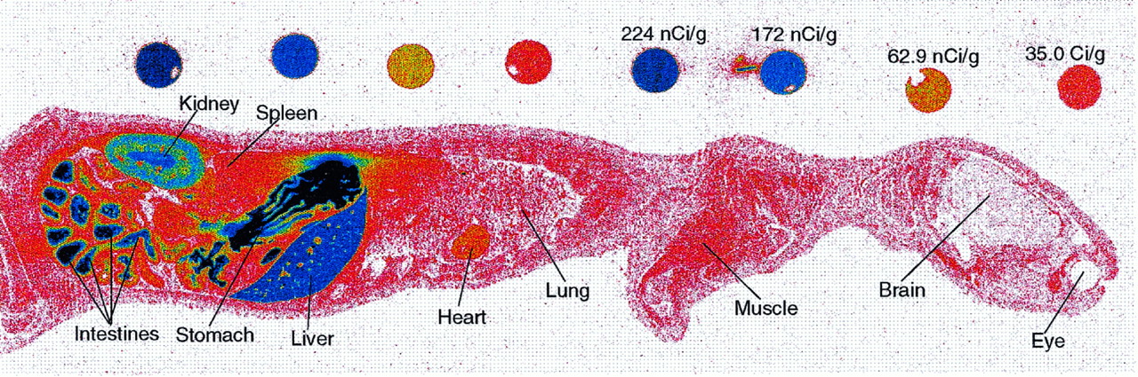
Representative whole body autoradioluminogram of a male rat at 1 hr following a single 10-mg/kg oral dose of 14C-labeled drug A.
Comparison of tissue concentrations of radioactivity in Sable Domestic male ferrets at 6, 16, and 30 min after oral administration of a single 10-mg/kg dose of 14C-labeled drug B4-a
Representative whole body autoradioluminogram of a male ferret at 16 min following a single 10-mg/kg oral dose of 14C-labeled drug B.
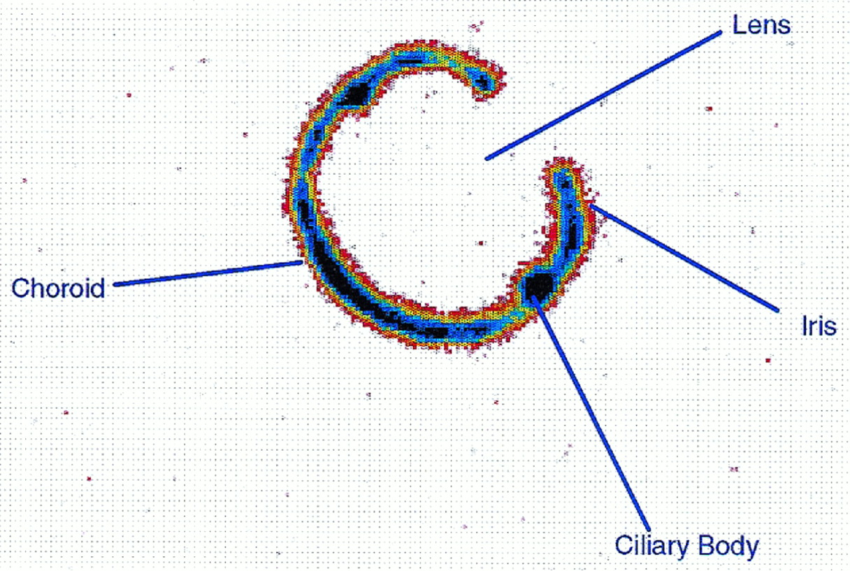
WBAL ocular radioactivity selectively partitioned in the melanin bearing regions of the rat eye. At 1 hr post-administration, WBAL uvea concentrations of drug A-associated radioactivity were the highest for all tissues examined except for alimentary tract contents. Uveal radioactivity (143 nCi/g) for the male rat was ca. 9.5 times higher than whole blood concentrations of 15 nCi/g. The choroid, ciliary bodies, and irises, which constitute the three major components of the uvea, showed differences in drug-related radioactivity at 1 hr post-dose for both the male and female rats. The radioactivity measured for the ciliary bodies was 2.9-fold (3.2-fold for female rat) higher than those levels detected for the irises. In contrast, choroid radioactivity was only 1.7-fold (1.5-fold for female rat) greater than iris radioactivity. The radioactivity present in these melanin-containing ocular tissues by 4 hr post-dose was slightly higher than the 1 hr concentrations for both male and female rats. The aqueous humor, lens, and vitreous body, which do not contain melanin, were devoid of radioactivity in both rat genders at 1 and 4 hr post-dose. DLSA evaluation of the intact right eye indicated a total radioactivity of 58 and 79 nCi/g and 10 and 46 nCi/g for the 1- and 4- hr male and female rats, respectively.
Concentrations of radioactivity measured by DLSA for male and female rat brain tissues at 1 and 4 hr post-dose confirmed WBAL evaluations that radioactivity present in the brain was below 5.9 nCi/g (table 3). The radioactivity concentration of 1.0 nCi/g present in ferret brain tissue at 6 min post-administration was below the WBAL lower quantitation limit of 5.9 nCi/g. At 16 and 30 min post-administration, WBAL and DLSA evaluations of ferret brain tissue radioactivity were similar.
Discussion
Attempts to develop calibration methods for quantitating the radioactive distribution of xenobiotics in tissues of animal whole bodies using a variety of calibration standards in conjunction with X-ray film as the medium to capture the image have been reported (Sainio and Sainio, 1991; Schweitzer et al., 1987; Somet al., 1983). Over- and underexposures are common when using X-ray film because the dynamic range is limited to less than 2 orders of magnitude. The reason for this is that calibration standards exposed to X-ray film produce a characteristic sigmoidal densityvs. exposure curve, which limits the dynamic range. A typical concentration range of 10 to 1000 nCi/g when using X-ray film requires multiple exposures of several days to several months to obtain quantitative information on the disposition of radiolabeled xenobiotics (Chay and Pohland, 1984). The use of inappropriate calibration standards and the lack of quality assurance controls (i.e.CQCS) have also provided unreliable WBA data (Irons and Gross, 1981;Ito and Brill, 1990). In this study, the standard curves prepared from loosely packed red blood cells were used to assess the accuracy and precision of autoradioluminography for determining the radioactivity content in tissues of whole bodies. The use of phosphorimaging screens, instead of X-ray film, provided excellent linearity in a single 4-day exposure that demonstrated a good correlation between the autoradioluminographic responses (Molecular Dynamic counts/μm2 minus background) and the radioactivity of each STD. The linear dynamic range spanned 5 orders of magnitude permitting the quantitation of both the high and low concentrations of radioactivity simultaneously from the same whole body cryosection in a single exposure. The use of CQCS as an assessment of cryosection thickness variability and overall WBAL assay performance allowed for the acceptance or rejection of those cryosections with coefficients of variations or relative errors not meeting predetermined quality assurance guidelines. Cryosections would be rejected if more than one-third of the calculated concentrations for the CQCS of any one cryosection varied from their nominal concentrations by more than 20%. Tissue concentrations from rejected cryosections would not be reported. Typically, accuracy and precision of cryosection CQCS vary by 3 to 7%. In addition, if the calculated concentrations of two-thirds of the CQCS at one concentration level varied by more than 20%, this cryosection would also be rejected.
STD and CQCS prepared from a tissue medium that closely resembles tissues under evaluation and sectioned at the same thickness (25 μm) as whole body cryosections eliminates the requirement for correction of sample thickness and self-absorption of β-particles in tissues of similar density. Inclusion of CQCS not only aids in the assessment of standard curve performance but also allows for the assessment of inter- and intra-cryosection variability. Establishment of such good laboratory practices for tissue distribution studies will successfully minimize the significance of cryosection thickness variations as the predominant source of variability and allow for reliable quantitation of radiolabeled xenobiotics in whole body cryosections. The results of this study demonstrated that the use of standard curve calibrators prepared from biological material, embedding CQCS in the specimen block, following a written validation protocol, and the establishment of quality assurance guidelines provided rapid and reliable data for the quantitative measurement of tissue radioactivity in whole body cryosections.
Both methods produced similar radioactivity concentrations for all tissues except for contralateral kidneys of one ferret and for rat ocular tissues. The right kidney of one ferret was the only tissue exhibiting different radioactivity levels between the two methods where a direct comparison was applicable. 14C-labeled drug B associated radioactivity in the right ferret kidney determined by DLSA was ca. 2-fold greater than that obtained from the left kidney by WBAL. Variations in renal blood flows, glomerular filtration rates, and rates of urinary elimination are factors that might have contributed to the 2-fold difference in radioactivity concentrations observed between the two contralateral kidneys of the same ferret.
As determined by WBAL, radioactivity present in rat ocular tissues was selectively partitioned only into the melanin impregnated tissues of the uveal tract. WBAL quantitation also revealed that the ciliary bodies contained substantially more radioactivity than the two other major uveal components: the choroid and irises. DLSA evaluation of specific ocular tissues was not plausible. Although DLSA evaluation of the intact eyeball revealed radioactivity was present in this organ, it was not possible to associate the location of this radioactivity to any specific ocular tissue. These observations demonstrate an advantage that WBAL has over radiometric methods by illustrating the quantitation and visualization of the heterogeneous localization of radioactivity in eye (fig. 4) and other tissues not readily available to dissection such as the intervertebral discs. The failure of the DSLA method to determine the localization of radioactivity in organ substructures, and the ability to do so by the WBAL method, clearly emphasizes the advantages in using the WBAL technique for tissue distribution studies.

Detail of a rat whole body autoradioluminogram emphasizing the uveal components of the eye.
A direct comparison between WBAL and DLSA could not be made when tissue radioactivity levels were less than the WBAL lower quantitation limit of 5.9 nCi/g. The lower limit of quantitation for DLSA for tissue radioactivity levels was primarily dependent upon the amount of tissue mass remaining after cryosectioning for sample oxidation. The minimal DLSA radioactive counting rate was established at 2 times background. Because tissue mass of each cryosection (25 μm) for a particular tissue in a given sectioning level should remain relatively constant, the autoradioluminographic lower limit of quantitation was dependent upon the quantity of radioactivity distributed to the target region and the linear response of β-particle energy transfer to photostimulatable phosphors (BaFBr:Eu+2). From the results of this study, it was quite apparent that WBAL provided accurate and precise quantitation of radioactivity in tissues of whole body cryosections that was comparable with DLSA evaluation of the same tissues. It was also evident that DLSA is more sensitive than WBAL, provided that ample amounts of tissue mass are available for analysis. However, increasing the cryosectioning thickness can improve WBAL sensitivity provided that self-absorption of14C-labeled β-particles does not become a limiting factor.
When comparing WBAL with DLSA, it is important to keep in mind that tissue concentrations of radioactivity could be greatly influenced by relatively high concentrations of radioactivity found in whole blood. Others investigators compared DLSA results obtained from exsanguinated animals to WBA results obtained from non-exsanguinated animals and thus may have unintentionally contributed to the perception that WBA was less precise than the dissect and radiometry methods. This source of error was eliminated in this study by using tissues obtained from the same specimen.
In conclusion, this study illustrated that the use of phosphorimaging for the quantitative analysis of 14C xenobiotics in tissues of whole body autoradioluminograms was comparable with the accuracy and precision of DLSA provided that radioactivity levels were above 5.9 nCi/g. WBAL quantitation of tissue radioactivity occurred over a wide dynamic range after a single, 4-day exposure. The advantage of using WBAL instead of DLSA for tissue distribution studies is realized by the comprehensive visualization and quantitation data of the distribution pattern for all tissues of the body. Thus, investigations involving discovery and developmental drugs are not limited to a finite number of preselected tissues. Localization of radioactivity in an unsuspected tissue as well as the heterogeneous distribution within a tissue may provide information pertaining to the metabolic fate, physiological response, or therapeutic effects of a drug. A primary objective for conducting an animal tissue distribution study is to project human tissue exposures to radioactivity required for conducting absorption, distribution, metabolism, and excretion studies in man. Thus, the sensitivity of WBAL is more than adequate to meet these dosimetry requirements, as the main concern is to predict the tissues that will be susceptible to high exposures to the radiolabeled drug.
Footnotes
-
Send reprint requests to: Michael J. Potchoiba, Department of Drug Metabolism, Central Research, Pfizer Inc., Eastern Point Road, Groton, CT 06340.
-
This study was presented during a poster session at the 1995 American Association of Pharmaceutical Scientists meeting (Abstract PPDM 8210).
- Abbreviations used are::
- DLSA
- dissection and liquid scintillation analysis
- WBA
- whole body autoradiography
- CQCS
- cryosection quality control samples
- STD
- standard curve calibrators
- QCS
- quality control samples
- WBAL
- whole body autoradioluminography
- Received July 3, 1997.
- Accepted November 24, 1997.
- The American Society for Pharmacology and Experimental Therapeutics


