


Abstract
Arylamine N-acetyltransferases (Nat) 1 and 2 catalyze the N-acetylation of aromatic amine and hydrazine drugs and carcinogens. After N-hydroxylation, they also catalyze the metabolic activation of N-hydroxy-arylamines via O-acetylation. Functional characterization of mouse Nat1 and Nat2 was investigated in an Nat2 knockout (KO) model and compared with the wild-type (WT) strain. Nat1- and Nat2-specific mRNA, determined by quantitative real-time polymerase chain reaction, was detected in all tissues examined and did not differ significantly (p > 0.05) between Nat2 KO and WT mice. Nat1 catalytic activity was present in all tissues examined and did not differ significantly (p > 0.05) between the Nat2 KO and WT mice. In contrast, Nat2 catalytic activity was present in all tissues examined from male WT mice but was below the limit of detection in all tissues of Nat2 KO mice. N-acetyltransferase activity toward the aromatic amine carcinogen 4-aminobiphenyl and O-acetyltransferase activity toward its proximate metabolite N-hydroxy-4-aminobiphenyl were both present in tissue cytosols of WT mice but were undetectable in Nat2 KO mice. Nat2 protein was readily detectable in liver cytosols of WT mice but not in liver cytosols from Nat2 KO mice. Since the reductions in Nat2 activity correlated with reductions in Nat2-specific protein but not mRNA, these results strongly suggest that insertion of the LacZ ablation cassette eliminated Nat2 protein and catalytic activity via disruption of the Nat2 protein, without significantly affecting transcription rates or transcript stability. The Nat2 KO model will be useful in future studies to assess the role of Nat2 in arylamine carcinogenesis.
Human N-acetyltransferases (NAT) 1 and 2 catalyze the N-acetylation of a variety of arylamine and hydrazine drugs and carcinogens (Hein et al., 2000; Butcher et al., 2002; Boukouvala and Fakis, 2005). In addition, after N-hydroxylation by cytochrome P450s, O-acetylation catalyzed by NAT1 and/or NAT2 leads to the bioactivation of N-hydroxylated metabolites generating unstable electrophilic intermediates capable of binding with nucleophiles such as DNA (Hein, 2002; Hein et al., 2006a,b) and potentially eliciting carcinogenic mutations.
Rodent models of the human NAT2 acetylation polymorphism have been reviewed (Levy et al., 1996; Hein et al., 1997, 2000; Boukouvala and Fakis, 2005). Three mouse Nat genes with 870-base pair intronless open reading frames were identified and designated as Nat1, Nat2, and Nat3 (Martell et al., 1992; Kelly and Sim, 1994). Later studies showed that all three mouse Nat genes colocalize on chromosome 8B3.1-3.3, a region syntenic with human 8p22 (Fakis et al., 2000; Boukouvala and Sim, 2005). The resulting 290-amino acid Nat proteins are highly analogous to NAT proteins in human and other mammalian species (Hein, 2002; Boukouvala and Fakis, 2005).
Nat2 exhibits genetic polymorphism in humans and mice. Variability in N-acetylation activity across mouse strains was shown by the observation that a few inbred mouse strains N-acetylated p-aminobenzoic acid (PABA) and benzidine at rates significantly lower than many other inbred mouse strains (Glowinski and Weber, 1982). Early evidence also supported the presence of a second locus, encoding for a second Nat isozyme (later identified as Nat1) with higher specificity for isoniazid (INH) (Hein et al., 1988). More recently, additional polymorphisms have been identified in mouse Nat2 from wild-derived inbred strains such as Mus spretus, which has seven single-nucleotide polymorphisms in the coding region, two of which are nonconservative leading to reductions in the hepatic Nat2 protein (Boukouvala et al., 2002). Partial purification of mouse hepatic Nat1 and Nat2 showed that INH is selective for catalysis by Nat1, whereas PABA is selective for catalysis via Nat2 (Hein et al., 1988). This selectivity was confirmed following recombinant expression of mouse Nat1 and Nat2 in COS-1 cells (Martell et al., 1992; Estrada-Rodgers et al., 1998).
Administration of 4-aminobiphenyl (ABP) to mice produces DNA adducts and tumors in urinary bladder and liver (Schieferstein et al., 1985; Dooley et al., 1992; Flammang et al., 1992; Poirier et al., 1995; McQueen et al., 2003a). Following recombinant expression of mouse Nat1 and Nat2 in Escherichia coli, ABP is N-acetylated by both Nat1 and Nat2 (Fretland et al., 1997). Metabolic activation (O-acetylation) of N-hydroxy-ABP by recombinant Nat1 and Nat2 has not been studied, but both Nat1 and Nat2 catalyze O-acetylation of N-hydroxy-2-aminofluorene (Fretland et al., 1997).
Various mouse models focusing on Nat2 have been reviewed (Levy et al., 1996; Boukouvala and Fakis, 2005). Among these various models, a murine Nat2 knockout (KO) model was created on a C57B1/6 background by the insertion of an ablative cassette into the Nat2 coding region, resulting in a nonfunctional gene product (Cornish et al., 2003). The objective of this study was to characterize Nat1 and Nat2 expression in this model and assess its utility for ABP carcinogenicity studies.
Materials and Methods
Tissue Procurement.Nat2 KO and WT mice were derived from colonies constructed and maintained at the University of Oxford. Procedures for their construction and an initial characterization have been reported previously (Cornish et al., 2003). Tissue samples were excised from adult male KO and WT mice (20–34 g each) immediately following cervical dislocation. Tissues were quickly removed (pancreas first to minimize autodigestion) rinsed with saline and snap-frozen in liquid nitrogen. The tissue samples were stored at –80°C at the University of Oxford prior to courier shipment on dry ice to the University of Louisville, where all subsequent procedures and measurements were performed.
Determination of RelativeNat1andNat2mRNA Levels. Relative levels of Nat1 and Nat2 mRNA normalized to β-actin mRNA were determined by quantitative real-time PCR. In brief, total RNA was extracted and purified using the Qiagen RNeasy Mini kit (Qiagen, Valencia CA), and reverse transcriptase (RT)-PCR was done using Superscript III (Invitrogen, Carlsbad, CA). Mouse Nat1- and Nat2-specific PCR primers and fluorogenic probes (Table 1) were designed using Primer Express (version 1.5; Applied Biosystems, Foster City, CA). Both forward and reverse Nat2-specific primers spanning positions 156 to 337 of the Nat2 coding region hybridized upstream of the site of insertion (position 534) of the ablation cassette, allowing the detection of an RNA species encoding N-terminal amino acids of Nat2 from wild-type and mutant alleles. The ABI 7700 sequence detection system (Applied Biosystems) was used to perform all PCR reactions in a total volume of 20 μl. Each reaction mixture contained 1× TaqMan Universal Master mix (Applied Biosystems), 300 nM each primer, and 100 nM probe. Five microliters of each cDNA (equivalent to 50 ng of reverse-transcribed total RNA) was used in each PCR, and experiments were carried out in triplicate. PCR conditions were initial incubation at 50°C for 2 min, followed by 10-min incubation at 95°C, and then 40 cycles of PCR at 95°C for 15 s and 60°C for 1 min. Each 96-well assay plate contained –RT and –cDNA controls.
PCR primers and TaqMan probes for quantitation of mouse Nat1- and Nat2-specific mRNA
Preparation of Tissue Cytosols. Individual organs from male Nat2 KO and WT mice were thawed on ice and homogenized (25% w/v) in 20 mM sodium phosphate, pH 7.4, containing EDTA (1 mM), dithiothreitol (1 mM), protease inhibitors aprotinin (1 μg/ml), phenylmethanesulfonyl fluoride (100 μM), and pepstatin (0.75 μM). Homogenates were centrifuged at 100,000g for 60 min at 4°C to prepare tissue cytosols. Cytosols were aliquoted and stored at –80°C until use.
Determination of Nat1 and Nat2 Catalytic Activity. Nat1 and Nat2 catalytic activity were measured with isoniazid (Hein et al., 1982, 1987b) and p-aminobenzoic acid (Leff et al., 1999), respectively, using methods described previously. In brief, suitably diluted tissue cytosol, 300 μM INH or PABA, and 1 mM acetyl coenzyme A were incubated at 37°C. Controls substituted water for acetyl coenzyme A. Acetyl-isoniazid or acetyl-p-aminobenzoic acid product was measured by absorbance at 303 or 280 nm, respectively.
ABP N-acetyltransferase assays were carried out as described previously (Fretland et al., 2002). In brief, reactions containing suitably diluted cytosol, ABP (1 mM), and acetyl coenzyme A (1 mM) were incubated at 37°C and terminated by the addition of 1/10 volume of 1 M acetic acid. ABP and N-acetyl-ABP were separated by reverse-phase high-performance liquid chromatography and quantitated by absorbance at 260 nm.
N-hydroxy-ABP O-acetyltransferase assays were carried out as described previously (Hein et al., 2006a) by measuring ABP-deoxyguanosine adduct levels that form spontaneously from reaction of unstable N-acetoxy-ABP with deoxyguanosine In brief, tissue cytosols were incubated with 1 mM N-hydroxy-ABP and acetyl coenzyme A and 1 mg/ml deoxyguanosine (made fresh daily) at 37°C. The ABP-deoxyguanosine adduct was separated by high-performance liquid chromatography and quantitated by absorbance at 300 nm.
Determination of Nat2 Protein Level. Nat2 protein was measured in liver cytosols from male Nat2 WT and KO mice as described previously (Zang et al., 2004) except for the antisera used. In brief, proteins were separated on a 12% Tris-glycine SDS-polyacrylamide gel (Cambrex Bio Science, Walkersville, MD), transferred to Hybond-ECL nitrocellulose membrane (Amersham Biosciences, Piscataway, NJ), and reacted with rabbit anti-mouse Nat2 (ES195), a polyclonal antiserum that, like a previously described antiserum (Stanley et al., 1996), was raised using the 12 C-terminal amino acids as hapten but complexed to soybean trypsin inhibitor as carrier. Chemiluminescent detection of Nat2 protein was achieved using SuperSignal West Pico Rabbit IgG Detection kit following the manufacturer's protocol (Pierce Biotechnology, Rockford, IL).
Results
Quantitative RT-PCR analysis was used to assess the relative level of Nat1 and Nat2 mRNA transcripts in different organs of Nat2 KO and WT male mice. No significant (p > 0.05) differences in relative Nat1 or Nat2 mRNA levels were observed between Nat2 KO and WT male mice in any tissue (Fig. 1). Nat1 catalytic activities, measured with the mouse Nat1-selective substrate isoniazid, were readily detectable in each organ of WT and KO mice. No significant differences (p > 0.05) in Nat1 catalytic activity were found between the Nat2 WT and KO mouse tissue (Fig. 2).
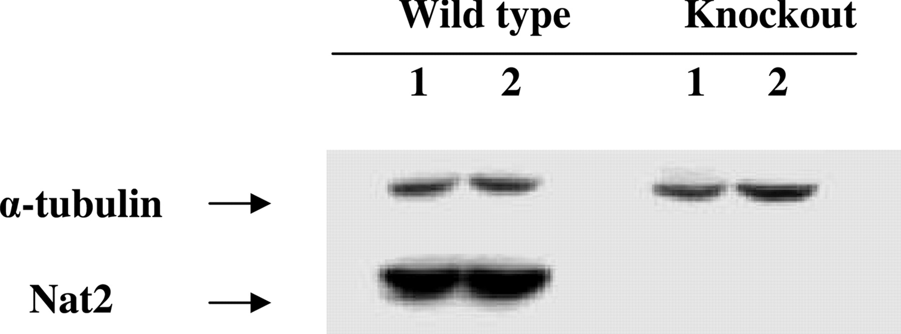
Nat2 catalytic activities, measured with mouse Nat2-selective substrate p-aminobenzoic acid, were readily detectable in each tissue from WT mice. In contrast, Nat2 catalytic activity levels were below the limit of detection in Nat2 KO mice (Fig. 3). Expression of Nat2 protein was readily observed in liver cytosol of WT mice but was below the limit of detection in Nat2 KO mice (Fig. 4).

Relative quantitation of mouse Nat1 (top) and Nat2 (bottom) mRNA levels in male WT and Nat2 KO mice. mRNA levels were determined using real-time quantitative real-time PCR normalized to β-actin mRNA levels. Bar, mean ± S.D. for three individual determinations on three different mice. Solid bars, WT mice; open bars, Nat2 KO mice. No significant (p > 0.05) differences in Nat1 or Nat2 mRNA levels were observed between WT and Nat2 KO mice in any tissue.

INH N-acetyltransferase activity in tissue cytosols of male Nat2 KO and WT mice. Solid bars, mean ± S.D. in WT mice; open bars, mean ± S.D. in Nat2 KO mice (n = 3). No significant (p > 0.05) differences in isoniazid N-acetyltransferase activities were observed between Nat2 KO and WT mice.
ABP N-acetyltransferase activities were readily detected in organ cytosols of WT mice but were below the limit of detection of Nat2 KO mice (Fig. 5). Likewise, N-hydroxy-ABP O-acetyltransferase activities were readily detected in tissue cytosols of WT mice but were below the limit of detection in Nat2 KO mice (Fig. 6).
Discussion
No significant (p > 0.05) differences in relative Nat1 or Nat2 mRNA levels were observed between Nat2 KO and WT male mice in any tissue (Fig. 1). The detection of both Nat1- and Nat2-specific transcripts in all hepatic and extrahepatic tissues examined of WT mice is consistent with previous studies in human (Windmill et al., 2000; Boukouvala and Sim, 2005) and mouse (Sugamori et al., 2003) tissues. In addition to the tissues studied here, mouse Nat2 transcript has been detected in mouse embryonic stem cells (Payton et al., 1999) as well as in postimplantation embryos throughout gestation (Mitchell et al., 1999; McQueen et al., 2003b). High levels of immunoreactive Nat2 protein have also been identified in the developing heart and neural tube of mouse embryos (Stanley et al., 1998), suggesting a possible endogenous role for mouse Nat2 in these tissues. Nevertheless, the Nat2 KO mice used in this study were healthy, and no pathologies were evident (Cornish et al., 2003). Likewise, no overt phenotype has been reported in Nat1/Nat2 double-knockout mice (Sugamori et al., 2003).

Expression of PABA N-acetyltransferase activity in tissue cytosols of male Nat2 KO and WT mice. Solid bars, mean ± S.D. from WT mice (n = 3). N-acetyltransferase activity was nondetectable (ND) in Nat2 KO mice. Limits of detection were approximately 6 pmol/min/mg in liver, 30 pmol/min/mg in gut, pancreas and urinary bladder, and 500 pmol/min/mg in prostate.

Western blot analysis of liver cytosols from wild type and Nat2 knockout mice. Hepatic cytosols (100 μg of total protein) from two wild-type and two Nat2 knockout mice were separated on 12% Tris-glycine SDS-polyacrylamide gel electrophoresis and probed with ES-195, a rabbit anti-mouse Nat2 antiserum.

Expression of ABP N-acetyltransferase activity in tissue cytosols of male Nat2 KO and WT mice. Solid bars, mean ± S.D. for ABP N-acetyltransferase activity from WT mice (n = 3). ABP N-acetyltransferase activity was nondetectable (ND) in Nat2 KO mice. Limits of detection were approximately 4 pmol/min/mg in liver and 20 pmol/min/mg in gut and pancreas.

Expression of N-hydroxy-ABP O-acetyltransferase activity in tissue cytosols of male Nat2 KO and WT mice. Solid bars, mean ± S.D. for N-hydroxy-ABP O-acetyltransferase activity from WT mice (n = 3). N-hydroxy-ABP O-acetyltransferase activity was nondetectable (ND) in Nat2 KO mice. Limits of detection were approximately 0.5 pmol/min/mg in liver, 1 pmol/min/mg in gut, and 5 pmol/min/mg in pancreas.
Since isoniazid is highly selective for mouse Nat1 (Hein et al., 1988), it was used to measure Nat1 catalytic activity in hepatic and extrahepatic tissues. Nat1 catalytic activity was readily detectable in each organ of WT and KO mice. No significant differences (p > 0.05) in Nat1 catalytic activity were found between Nat2 WT and KO mouse tissues (Fig. 2). Neither a reduction nor compensatory enhancement in Nat1 catalytic activity or mRNA were observed in the Nat2 KO mice, a result consistent with previous studies (Cornish et al., 2003). It was expected that mouse Nat1 catalytic activity would either be solely or predominantly expressed in liver and gut. Thus, a particularly novel finding of our study is that mouse Nat1 catalytic activity, as measured by INH N-acetylation, and mRNA transcript were present not only in liver and gut but also in extrahepatic mouse tissues. Furthermore, the Nat1 catalytic activities were fairly uniform across the hepatic and extrahepatic (gut, pancreas, urinary bladder, and prostate) tissues examined. Recently, similar evidence for extrahepatic distribution of Nat1 catalytic activity was observed in tissue cytosols of Syrian hamsters (Hein et al., 2006a) and rats (M. A. Doll, J. R. Neale, J. Bendaly, and D. W. Hein, unpublished data) congenic for Nat2. The possibility exists that extrahepatic INH N-acetyltransferase activity was catalyzed by mouse Nat2 rather than Nat1. However, the INH N-acetyltransferase activities were equivalent in the WT and Nat2 KO mice, providing strong evidence that the activity was catalyzed specifically by mouse Nat1 in each tissue and not by Nat2.
Nat2 catalytic activity, measured with mouse Nat2-selective substrate p-aminobenzoic acid, was readily detectable in each tissue from WT mice. In contrast, Nat2 catalytic activity levels were below the limit of detection in Nat2 KO mice (Fig. 3). Nat2 catalytic activities in the wild-type mice were fairly uniform across the hepatic and extrahepatic tissues (gut, pancreas, urinary bladder, and prostate) examined in this study. This finding is consistent with previous studies showing that mouse Nat2 expression (Stanley et al., 1997; Cornish et al., 2003) and catalytic activity (Chung et al., 1993; Payton et al., 1999) are widely distributed. Nat2 catalytic activity also has been shown to be widely distributed in the Syrian hamster (Hein et al., 1987a,b, 1991b, 2006a) and rat (Hein et al., 1991a). As expected, Nat2 catalytic activity in each of these tissues was substantially reduced (below the level of detection) in the same hepatic and extrahepatic tissues of the Nat2 KO mice. The reduction in Nat2 catalytic activity was reflected in a substantial reduction in Nat2 protein (Fig. 4) but was not secondary to reductions in Nat2-specific mRNA (Fig. 1) in the Nat2 KO mice. Since the reductions in Nat2 activity correlated with reductions in Nat2 protein but not mRNA, these results strongly suggest that insertion of the LacZ ablation cassette eliminated Nat2 protein and catalytic activity via disruption of the Nat2 protein, without significantly affecting transcription rates or transcript stability.
ABP N-acetyltransferase activity was readily detected in organ cytosols of WT mice but were below the limit of detection of Nat2 KO mice (Fig. 5). Likewise, N-hydroxy-ABP O-acetyltransferase activities were readily detected in tissue cytosols of WT mice but were below the limit of detection in Nat2 KO mice (Fig. 6). Since previous studies have shown that ABP is N-acetylated by both mouse Nat1 and Nat2 (Fretland et al., 1997), we were surprised to observe that both the N-acetylation of ABP and O-acetylation of N-hydroxy-ABP were undetectable in Nat2 KO mouse tissues despite the fact that Nat1 catalytic activity (measured as INH Nat catalytic activity) was readily expressed in these tissues. The molecular basis for this observation is unknown, but these results are consistent with findings in rapid and slow acetylator Syrian hamsters (Hein et al., 2006a) and rats (M. A. Doll, J. R. Neale, J. Bendaly, and D. W. Hein, unpublished data) congenic for Nat2. Thus, Nat2 is important in both the inactivation (N-acetylation) and activation (O-acetylation) of ABP in the mouse, and future ABP bioassays in WT and Nat2 KO mice should be very useful in further investigating the role of Nat2 in aromatic amine-induced toxicity and carcinogenesis.
Footnotes
-
This work was supported by research grants from the Wellcome Trust and the University of Louisville Summer Research Scholars Program and by United States Public Health Service Grant R01-CA34627 from the National Cancer Institute. A preliminary report of this work was presented at the Annual Meeting of the Society of Toxicology (Abstract no. 1266); 2005 March 6–10; New Orleans, LA. Society of Toxicology, Reston, VA.
-
Article, publication date, and citation information can be found at http://jpet.aspetjournals.org.
-
doi:10.1124/jpet.106.108662.
-
ABBREVIATIONS: NAT, human N-acetyltransferase; Nat, mouse N-acetyltransferase; PABA, p-aminobenzoic acid; INH, isoniazid; ABP, 4-aminobiphenyl; KO, knockout; WT, wild type; PCR, polymerase chain reaction; RT, reverse transcriptase.
- Received May 30, 2006.
- Accepted July 19, 2006.
- The American Society for Pharmacology and Experimental Therapeutics


