


Abstract
Treatment of rats with peroxisome proliferators is known to affect gene expression, including suppression of CYP2C11. The current study examined the mechanism of negative regulation of CYP2C11, comparing the effects of a classic peroxisome proliferator, nafenopin, with those of the steroid dehydroepiandrosterone (DHEA). In vivo dose-response experiments for DHEA were carried out with rats. Only the highest dose of DHEA in the diet (0.45%), a dose previously shown to produce peroxisome proliferation, caused suppression of CYP2C11 expression. Lower doses of DHEA (0.012 to 0.20% in diet) had little effect on CYP2C11 expression. In HepG2 cells, negative regulation of a CYP2C11 reporter gene by nafenopin required coexpression of PPARα, whereas negative regulation by DHEA did not. Deletion analysis revealed that the responsive region for both DHEA and nafenopin was between -108 and -60 relative to the transcription start site. Mutations in several putative transcription factor binding sites in the 5′-flanking region of CYP2C11 were produced. A mutation at -121 bp significantly diminished basal expression of CYP2C11 but did not affect negative regulation by DHEA or nafenopin. A mutation at -75 bp had only a small effect on basal expression but completely abolished negative regulation by DHEA and nafenopin. Gel shift experiments indicated that PPARα/RXRα heterodimers do not bind DNA in this region. Therefore, the sequence at -75 bp of CYP2C11 is necessary for negative regulation by both DHEA and nafenopin. However, the upstream events leading to suppression at this site must differ for DHEA and nafenopin.
The purpose of the current work was to examine the mechanism of suppression of CYP2C11 and to compare the effects of dehydroepiandrosterone (DHEA) with those of the classic peroxisome proliferator, nafenopin. Peroxisome proliferators are structurally diverse compounds that activate the peroxisome proliferator-activated receptor α (PPARα), a transcription factor that modulates expression of many genes. Peroxisome proliferators include fibric acid derivatives, fatty acids, arachidonic acid metabolites, and phthalate ester plasticizers (Issemann and Green, 1990; Göttlicher et al., 1992;Yu et al., 1995). PPARα is a member of the nuclear receptor superfamily of ligand-activated transcription factors and functions as an obligate heterodimer with the retinoid X receptor α (RXRα). PPARα/RXRα heterodimers can activate transcription of a wide array of genes, including those coding for proteins involved in lipid metabolism such as acyl CoA oxidase and cytochrome P450 4A. PPARα/RXRα heterodimers bind to sequences of DNA containing direct repeats of the hexanucleotide sequence AGGTCA separated by one nucleotide, a DR1 motif (IJpenberg et al., 1997). PPARα/RXRα heterodimers can also modulate gene expression via DNA-binding independent mechanisms through protein-protein interactions with other transcription factors and coactivator proteins (Delerive et al., 1999).
Peroxisome proliferators are so-named because in response to these chemicals, rodents develop a pathology of the liver characterized by an increase in size and number of peroxisomes, increased expression of peroxisomal, mitochondrial, and microsomal enzymes, and hepatomegaly (Reddy and Chu, 1996). The peroxisome proliferative response does not occur in humans, either because human PPARα is expressed at relatively low levels compared with rodent PPARα or because human liver lacks a component of the transduction pathway necessary for PPARα-mediated effects (Hertz and Bar-Tana, 1998; Palmer et al., 1998).
DHEA is a naturally occurring steroid hormone in humans. It is secreted by the adrenal gland, largely as the sulfate conjugate, and is the most abundant circulating steroid in humans. DHEA-sulfate is taken up by target tissues, converted to DHEA by the action of sulfatases, and DHEA is then converted to many other steroid hormones, such as androstenedione, androstenediol, testosterone, and estradiol (Kroboth et al., 1999).
Supplementation with exogenous DHEA has many purported health benefits in humans, including chemoprotection, enhancement of immune function, improvements in mood and energy, and antiobesity effects (Kroboth et al., 1999). DHEA is widely available over the counter and is considered a dietary supplement. Treatment of rodents with DHEA produces many beneficial effects, including chemoprevention in multiple cancer models (Lubet et al., 1998), anti-obesity effects (Yen et al., 1977), amelioration of diabetes and systemic lupus erythematosus (Coleman et al., 1982), and enhancement of immune function (Araneo et al., 1995). However, treatment of rodents with high doses of DHEA produces peroxisome proliferation (Wu et al., 1989). The modulation of gene expression by DHEA is typical of other peroxisome proliferators (Yamada et al., 1991). The in vivo induction of peroxisome proliferator-responsive genes by DHEA is dependent on PPARα, as shown in experiments using PPARα-null mice. DHEA-sulfate induces a battery of peroxisome proliferator responsive genes in wild-type mice but is unable to induce these genes in PPARα-null mice (Peters et al., 1996). However, DHEA differs from the classic peroxisome proliferator chemicals in that it is apparently not a ligand for PPARα (Krey et al., 1997) and it is unable to activate PPARα in in vitro assays (Peters et al., 1996; Waxman, 1996). The mechanism of activation of PPARα by DHEA is not known at this time.
In addition to the well characterized ability of peroxisome proliferators to induce expression of many genes, these compounds can also suppress gene expression, including the genes for transferrin, apolipoprotein CIII, and CYP7A1 (Hertz et al., 1995, 1996; Marrapodi and Chiang, 2000). There is evidence that suppression may require PPARα activation through both DNA-binding-dependent and -independent mechanisms (Hertz et al., 1995; Delerive et al., 1999; Marrapodi and Chiang, 2000). Additionally, there is evidence for PPARα-independent mechanisms for suppression of gene expression by fibrates (Staels et al., 1995).
One of the genes suppressed by peroxisome proliferator chemicals is cytochrome P450 2C11 (CYP2C11). CYP2C11 is expressed in male rat liver and codes for a protein involved in hydroxylation of steroids, epoxygenation of arachidonic acid, and drug metabolism. There are related genes in humans that code for proteins involved in steroid hydroxylation reactions, CYP2C9 and CYP2C19. Whether peroxisome proliferator chemicals suppress expression of human CYP2C genes is not known. However, expression of CYP2C11 is markedly suppressed by peroxisome proliferator chemicals (Corton et al., 1998) and by DHEA (Prough et al., 1995).
Materials and Methods
Chemicals. DHEA and DHEA-sulfate were purchased from Steraloids (Newport, RI) and nafenopin was a gift from Novartis (Ardsley, NY). Ciprofibrate was purchased from Sigma (St. Louis, MO). Oligonucleotides were purchased from Operon Technologies (Alameda, CA).
Animal Treatments. Male Sprague-Dawley rats (180–200 g; Hsd:SD, Harlan, Indianapolis, IN) were acclimated to control diet (AIN-76A; ICN Biomedicals, Cleveland, OH) for 3 days before treatment. Animals (three or four per treatment group) were maintained on control diet alone or diet supplemented with 0.012, 0.06, 0.2, or 0.45% DHEA for 5 days. Diets containing DHEA were generously provided by Clinton Grubbs (University of Alabama at Birmingham). An additional group of animals was maintained on control diet and given daily i.p. injections of nafenopin (30 mg/kg in corn oil) for four days. Animals were sacrificed and livers were perfused with 0.9% saline, excised, and immediately frozen in liquid nitrogen and stored at -80°C.
Western Blot Analysis. The levels of CYP2C11 protein in rat liver microsomal fractions were measured as described by Wu et al. (1989) using an antibody against CYP2C11 obtained from Oxford Biomedical (Oxford, MI). The protein-antibody complex formed was quantified using the ECF chemofluorescent detection system (Amersham Biosciences, Piscataway, NJ) with a Molecular Dynamics Storm 860 PhosphorImager (Amersham Biosciences).
Northern Blot Analysis. Total RNA was isolated from livers using guanidinium hydrochloride/phenol extraction with TRIzol reagent (Invitrogen, Carlsbad, CA). RNA (12 μg) was separated by electrophoresis on a 1% agarose/10% formaldehyde gel and transferred to ζ-probe nylon membranes (Bio-Rad, Hercules, CA). Plasmids containing cDNA for rat CYP4A1 and rat glyceraldehyde 3-phosphate dehydrogenase were generously provided by James Hardwick (Northeastern Ohio Universities College of Medicine, Rootstown, Oh), and Jean-Marie Blanchard (University des Sciences et Techniques du Languedoc, Montpellier, France), respectively. A 412-bp cDNA probe for CYP2C11 was generated by reverse transcription-PCR of male rat liver RNA using the primers 5′-tctgcttctcctctcactct-3′ and 5′-ctgagcctcctcttgaatac-3′. The PCR product was subcloned into pCR2.1 and sequenced. Probe labeling, hybridizations, and washes were carried out as described previously (Ripp et al., 2002). Blots were exposed to phosphor screens and analyzed using a PhosphorImager and ImageQuant software (Amersham Biosciences).
Plasmids. The CYP2C11 reporter plasmid 1300/2C11-Luc was constructed by excising the 1300-bp HindIII/BamHI fragment from 1300/2C11-CAT (Ström et al., 1994) and ligating it into a modified pGL3-basic vector, pSLR1. pSLR1 was constructed by filling in the BamHI and SalI sites of pGL3-basic (Promega), then inserting a linker into the HindIII/BglII digested plasmid. The linker contained BamHI and SalI sites, in that order. The reporter plasmids 348/2C11-Luc, 143/2C11-Luc, 108/2C11-Luc, and 60/2C11-Luc were constructed by digesting the respective pGL2-based plasmids (Ström et al., 1994) with KpnI and PstI and ligating the isolated fragments into a KpnI/PstI-digested pSLR1. The reporter plasmid 563/2C11-Luc was constructed by digesting 1300/2C11-Luc with KpnI and ligating the vector. The reporter plasmid 197/2C11-Luc was constructed by digesting 348/2C11-Luc with KpnI and SanDI and filling in and ligating the vector fragment. All CYP2C11 luciferase constructs described above extend to +25 of the CYP2C11 structural gene relative to the transcription start site. The luciferase reporter plasmid GSTmin was constructed by isolating the GSTA2 minimal promoter from 0.164YaLUC (Falkner et al., 1998) and inserting it into the HindIII/KpnI sites of pGL3-basic. The luciferase reporter plasmid PPRELuc contains two copies of the peroxisome proliferator responsive element from rat fatty acyl CoA oxidase upstream of the promoter of GSTmin. The β-galactosidase expression vector, pCMVβ, was purchased from BD Bisociences Clontech (Palo Alto, CA). The expression vectors for murine PPARα and human RXRα were kindly provided by Thomas Rushmore (Merck Research Laboratories, West Point, PA) and Dan Noonan (University of Kentucky, Lexington, KY), respectively.
Mutagenesis of CYP2C11 5′-Flanking Region. The wild-type plasmid 1300/2C11-Luc was used as the template to generate mutated sequences by overlap extension PCR (Ho et al., 1989). Complementary internal primers containing mutations as well as wild-type primers upstream in 1300/2C11-Luc and downstream in pGL3-basic were used. The external primers were 5′-cttagttggctcatgctgagg-3′ (CYP2C11 upstream primer) and 5′-tttctttatgtttttggcgtcttcca-3′ (pGL3 downstream primer). The internal primers containing mutations were as follows (mutations underlined): mut302, 5′-ggttagagatctgtcagaggctagtca-3′ and its complementary sequence; mut121, 5′-attttaacacctcagggtccacaaag-3′ and its complementary sequence; mut75, 5′-gttgattcctcacttaggtatcagaa-3′ and its complementary sequence; mut66, 5′-gttgattggtcacttacctatcagaa-3′ and its complementary sequence. Final PCR products were subcloned into pCR2.1 (Invitrogen) and sequenced to determine orientation. Correctly oriented CYP2C11 sequences containing mutations were excised from pCR2.1 using KpnI/BamHI and cloned into a KpnI/BamHI-digested 563/2C11-Luc. All final plasmids were sequenced to verify the presence of the mutations.
Cell Culture and Transient Transfection Assays. CV-1, H4IIE, and HepG2 cells (American Type Culture Collection, Manassas, VA) were cultured in Dulbecco's modified Eagle's medium supplemented with 10% fetal bovine serum and penicillin/streptomycin/amphotericin B. Cells were grown in a 37°C, 5% CO2, humidified atmosphere and subcultured every 2 to 3 days. For transient transfection experiments, cells were plated in 12-well plates at a cell density of 1.5 × 10⁵ cells per well (CV-1 and HepG2) or 1.0 × 10⁵ cells per well (H4IIE) 18 to 24 h before transfection. CV-1 and HepG2 cells were transfected using LipofectAMINE (2 μl per well; Invitrogen) and H4IIE cells were transfected using Superfect (2.5 μl per well; QIAGEN, Valencia, CA), according to the manufacturer's instructions, under serum-free conditions. Cells were typically transfected with CMVβ (0.5 μg per well), reporter vector (1 μg per well for CYP2C11 constructs, 0.25 μg per well for other reporter vectors), and expression vector (0.1 μg per well). Twenty-four hours after transfection, medium was changed, and cells were treated with 500× stocks of DHEA or nafenopin in dimethyl sulfoxide, for a final treatment concentration of 35 μM. Higher levels of DHEA have been shown to deplete ribo- and deoxyribonucleoside pools and inhibit tumor cell growth and limit transcription (Garcea et al., 1988). Cells were then harvested after 24 h using 100 μl of cell lysis reagent (Promega, Madison, WI). Luciferase assays (using 20 μl of cell extract) and β-galactosidase assays (using 5 μl cell extract) were carried out as described previously (Falkner et al., 1998). Transfection experiments were conducted in triplicate or quadruplicate and means ± standard deviations were determined. Statistical comparisons among treatment groups were determined using a two-tailed t test, with p < 0.05 as the criterion for significance. Experiments were repeated at least twice to confirm results.
Electrophoretic Mobility Shift Assays. Double-stranded DNA probes were generated by annealing complementary oligonucleotides that contained 5′ overhangs. The sequence of the positive control probe, containing the rat fatty acyl CoA oxidase PPRE, was 5′-gtacgggaccaggacaaaggtcacgttcg-3′. The sequence for two CYP2C11 peroxisome proliferator responsive regions were 5′-gtac-atctagttgattggtcacttaggtatcagaa-3′ (-86 to -56 of CYP2C11) and 5′-gtacagcatatctagttgattggtcactt-3′ (-91 to -67 of CYP2C11). A longer CYP2C11 probe was generated by digesting 563/2C11-Luc with BamHI/SanDI and gel-purifying the 225-bp fragment containing -198 to +25 of CYP2C11. An additional probe was generated by digesting 348/2C11-Luc with KpnI/SanDI and gel-purifying the fragment containing -348 to -198 of CYP2C11. All probes were radiolabeled by filling in 5′ overhangs using Klenow DNA polymerase and 32P-labeled dATP or dTTP. PPARα and RXRα were generated by in vitro transcription and translation of 1 μg of plasmid DNA using a commercially available kit (Promega, Madison, WI). Initially, in vitro transcription and translation reactions were carried out in the presence of 35S-labeled methionine. Reactions were resolved by SDS-polyacrylamide gel electrophoresis, and the presence of protein of the expected molecular weight was verified. For electrophoretic mobility shift assay experiments, 4 μl of transcription/translation reactions were incubated with poly dIdC (1 μg), binding buffer (11 mM HEPES, pH 7.9, 50 mM KCl, 1 mM dithiothreitol, 2.5 mM MgCl2, and 10% glycerol), and water to a volume of 19 μl. In some cases, unlabeled probe was added to the binding reactions, and reactions were incubated on ice for 20 min. Radiolabeled probe (1 pmol) was added and reactions were incubated at room temperature for 20 min. DNA-protein complexes were resolved on 4% polyacrylamide gels run in 0.5× Tris-borate/EDTA buffer. Gels were dried and analyzed by PhosphorImager analysis.
Results
CYP2C11 Is Negatively Regulated by Peroxisome Proliferators in Vivo. CYP2C11 mRNA and enzymatic activity have previously been shown to be negatively regulated by DHEA (Prough et al., 1995) and by the classic peroxisome proliferators WY-14,643, gemfibrozil, and di-n-butyl phthalate (Corton et al., 1998). Previously, only the effects of a high dose of DHEA had been examined. Therefore, initial studies were directed at determining the effect of a range of DHEA doses. Rats were treated for 5 days with control diet or diet supplemented with 0.012, 0.06, 0.2, or 0.45% DHEA (these doses are equivalent to 6, 30, 100, and 225 mg/kg per day, respectively, assuming an average food intake of 10 g per day). An additional group of rats received control diet plus daily i.p. injections of nafenopin (30 mg/kg), a dosage we have observed to give maximal induction of CYP4A1. Total RNA was isolated from liver and northern analyses were performed (Fig. 1). As a positive control, mRNA levels of the peroxisome proliferator-responsive CYP4A1 were determined. CYP4A1 was induced by DHEA in a dose-dependent manner. As expected, nafenopin also induced CYP4A1. For CYP2C11, lower doses of DHEA produced a slight increase in mRNA levels, whereas the highest dose produced a decrease in mRNA levels. Nafenopin treatment caused a decrease in CYP2C11 mRNA, as expected based on previous studies (Prough et al., 1995). Western blot analysis of liver microsomal fractions from untreated and DHEA fed rats demonstrated that DHEA decreased the levels of CYP2C11 protein by 60% at the highest dose (data not shown). As was shown for the peroxisome proliferator di-n-butylphthalate by Corton et al. (1998), nafenopin did not significantly alter the level of CYP2C11 protein, although CYP4A1 was induced by nafenopin (data not shown).

Dose-response for regulation of rat hepatic CYP2C11 and CYP4A1 by DHEA treatment in vivo. Male Sprague-Dawley rats were treated for 5 days with control diet, diet supplemented with the indicated concentrations of DHEA, or control diet plus daily i.p. injections of 30 mg/kg nafenopin. Liver RNA was subjected to Northern analysis using 32P-labeled cDNA probes for CYP2C11, CYP4A1, and glyceraldehyde 3-phosphate dehydrogenase. A, PhosphorImages of representative experiment. B, quantitation of PhosphorImages. Data are expressed as percentage control ± S.D. *, p < 0.05, significantly different from control.
Activation of Gene Expression by Peroxisome Proliferators Can Be Reproduced in Transiently Transfected HepG2, CV-1, and H4IIE Cells. To elucidate the mechanism of negative regulation of CYP2C11 by DHEA and nafenopin, an in vitro model for CYP2C11 negative regulation was developed. Several cell lines were tested for their ability to mediate peroxisome proliferator responsiveness. HepG2 (human hepatoblastoma), CV-1 (monkey kidney), and H4IIE (rat hepatoma) cell lines were transiently transfected with luciferase reporter constructs containing the minimal promoter from the glutathione S-transferase A2 gene (GSTmin), or two copies of the peroxisome proliferator responsive element (PPRE) from the rat fatty acyl CoA oxidase gene in front of the minimal promoter from the GSTA2 gene (PPRELuc). Cells were cotransfected with either pSG5, PPARα, RXRα, or a combination of receptors and treated with the fibric acid peroxisome proliferator, nafenopin, or with DHEA. As shown in Fig. 2, all cell lines tested were capable of supporting activation of the PPRE reporter in response to nafenopin. In HepG2 and H4IIE cells, very little (<1.5-fold) activation of the reporter occurred in the absence of cotransfected PPARα expression vector, indicating that these cell lines express little endogenous PPARα. However, CV-1 cells apparently express endogenous PPARα, as indicated by the 3.5-fold induction of PPRELuc activity in the absence of cotransfected PPARα expression vector.

Peroxisome proliferator responsiveness can be reproduced in transient transfection assays in multiple cell lines. HepG2 (A), H4IIE (B), and CV-1 (C) cell lines were transfected with either PPRELuc or GSTmin reporter plasmids (0.25 μg/well), the indicated expression vector (100 ng/well), and CMV-β (0.5 μg/well) as described under Materials and Methods. Empty expression vector (pSG5) was used to equalize DNA concentrations. Twenty-four hours after transfection, cells were treated with DMSO (vehicle control), nafenopin (25 μM), or DHEA (25 μM) and harvested 24 h later. Data represent mean ± S.D. for triplicate wells and are expressed relative to GSTmin activity in the absence of coexpressed receptor. **, p < 0.05, significantly different from PPRELuc in the absence of coexpressed receptor. *, p < 0.05, significantly different from DMSO treatment.
Cotransfection of PPARα expression vector increased basal expression of the PPRELuc reporter and enhanced induction by nafenopin in all cell lines tested. The enhanced expression of the reporter when PPARα is cotransfected, even in the absence of added ligand, is a phenomenon that has been noted previously with PPARα and other nuclear receptors in cell-based assays (Peters et al., 1996; Ripp et al., 2002). This phenomenon may be caused by a ligand-independent activation (e.g., via phosphorylation) of the transfected receptors or might be caused by ligands present in the cell culture medium or serum.
Cotransfection of an expression vector for RXRα was not required for PPARα-dependent activation, indicating that these cell lines express sufficient endogenous RXRα to support PPARα-dependent responses. Cotransfection of expression vector for RXRα in addition to PPARα markedly increased basal expression of the reporter construct, which resulted in diminished fold-activation by nafenopin. The ability of DHEA (25 μM) to activate the PPRE-containing reporter was also tested in the three cell lines. DHEA either had no effect (H4IIE cells) or had a small inhibitory effect (CV-1 and HepG2 cells) on both PPRELuc and the minimal GST promoter (Fig. 2). DHEA-S (25 and 50 μM) was also tested and had no effect on expression in any of these cell lines (data not shown). These results indicate that although DHEA and DHEA-S activate peroxisome proliferator-responsive genes in vivo, they are not able to produce this response in vitro, even though classic peroxisome proliferators, such as nafenopin, can activate PPARα in vitro. This is in agreement with other investigations that have tested DHEA and DHEA-S in various cell-based assays (Peters et al., 1996; Waxman, 1996).
Peroxisome Proliferator-Dependent Negative Regulation of CYP2C11 Can Be Reproduced in Transiently Transfected HepG2 Cells. To elucidate the mechanism of negative regulation of CYP2C11 by peroxisome proliferators, a CYP2C11 reporter construct was transiently transfected into the HepG2, CV-1, or H4IIE cell line. This reporter plasmid contained the promoter and 5′-flanking region (-1287 to +25 relative to the transcription start site) of the CYP2C11 gene upstream of a luciferase reporter gene (designated 1300/2C11-Luc). 1300/2C11-Luc was expressed at readily detectable levels in HepG2 cells and CV-1 cells, but expression was very low in H4IIE cells (data not shown). It should be noted that there was some variability in the ability of HepG2 cells to maintain basal expression of the CYP2C11 reporter. At times, basal expression was readily detectable above background; at other times, basal expression was very low. Variability in apolipoprotein CIII secretion by HepG2 cells has been reported, and this variability was attributed to factors in the serum used in the culture medium (Clavey et al., 1999). Various culture conditions were examined to try to explain the variability in CYP2C11 expression, but the reason for this variability remains unknown. Preliminary experiments indicated that negative regulation of the CYP2C11 reporter could be reproduced in HepG2 cells but not in CV-1 cells (data not shown). Therefore, subsequent experiments were carried out with subcultures of HepG2 cell line, which we observed to be growing rapidly. When HepG2 cell cultures were growing slowly, the variability in expression of CYP2C11 constructs was much more pronounced.
The reporter plasmid 1300/2C11-Luc was transiently transfected into HepG2 cells, in either the absence or the presence of coexpressed PPARα. Cells were then treated with DMSO (vehicle control), or 35 μM DHEA, nafenopin, ciprofibrate, or DHEA-sulfate (Fig. 3). In the absence of coexpressed PPARα, only DHEA had an effect on CYP2C11 reporter activity, suppressing expression by approximately 40% (Fig. 3A). When PPARα was coexpressed, basal expression of the CYP2C11 reporter decreased by approximately 50% (compare DMSO treatment in Fig. 3, A versus B). This is consistent with the ligand-independent activation of PPARα noted above for the transactivation assay. Treatment with nafenopin, ciprofibrate, or DHEA produced an additional 50% suppression of reporter activity (Fig. 3B). These results indicate that negative regulation of CYP2C11 by peroxisome proliferators can be reproduced in transiently transfected HepG2 cells. The classic peroxisome proliferators nafenopin and ciprofibrate required PPARα, whereas DHEA suppressed expression in a PPARα-independent manner.

Coexpression of PPARα is required for negative regulation of CYP2C11 by nafenopin and ciprofibrate but not by DHEA. HepG2 cells were transfected with 1300/2C11-Luc reporter plasmid (1 μg) and pCMVβ control plasmid (0.5 μg), ± PPARα expression vector (0.1 ug) as described under Materials and Methods. Cells were treated with 35 μM concentrations of the indicated compounds for 24 h. Data represent the mean ± S.D. of four wells. *, p < 0.05, significantly different from DMSO-treated controls.
Concentration-Dependence of the CYP2C11 Reporter Expression to DHEA and Nafenopin in HepG2 Cells. To compare the potency of DHEA and nafenopin with regard to negative regulation of CYP2C11 in vitro, concentration-response analyses were carried out in HepG2 cells. Nafenopin suppressed reporter gene expression at concentrations between 1 and 50 μM, whereas DHEA required slightly higher concentrations, between 10 and 50 μM (Fig. 4). However, higher concentrations of both DHEA and nafenopin (>50 μM) also suppressed expression of the parent vector pSLR1 (Fig. 4). This suppressive effect of DHEA at concentrations greater than 50 μM was also noted with luciferase reporter vectors containing the glutathione S-transferase A2 minimal promoter and the human quinone oxidoreductase minimal promoter, but not with the CMV promoter (data not shown). This suppressive effect is probably not caused by cell toxicity, because the expression of CMV promoter-driven vectors was not affected and overall cellular protein levels were not altered by DHEA (data not shown). The suppressive effect of DHEA on CYP2C11 expression occurred at lower concentrations than with other promoters, but at higher DHEA concentrations, the suppressive effect of DHEA was not specific to CYP2C11. Therefore, subsequent experiments were carried out with DHEA and nafenopin concentrations of 35 μM, conditions under which the parent vector was not affected.

Concentration-response for suppression of reporter activity. HepG2 cells were transfected with either 1300/2C11-Luc or pGL3-basic reporter vectors (1 μg/well) and expression vector for PPARα (0.1 μg/well). Twenty-four hours after transfection, cells were treated with varying concentrations of nafenopin (NAF) or DHEA and harvested 24 h later. Data are expressed relative to untreated cells, mean ± S.D.
Results of these in vitro experiments suggested that negative regulation of CYP2C11 by DHEA was not dependent on PPARα. Therefore, an attempt was made to conclusively assess this by determining regulation by DHEA in wild-type versus PPARα-null mice. PPARα (+/+) and (-/-) mice were treated with DHEA-sulfate, and effects on hepatic enzyme expression were assessed, as described previously (Ripp et al., 2002). Unfortunately, the mouse Cyp2c29 gene, thought to be the homolog to rat CYP2C11, was not regulated by DHEA treatment in either wild-type or PPARα (-/-) animals (data not shown). Therefore, these in vivo experiments were inconclusive with regard to the requirement for PPARα in CYP2C11 regulation.
5′-Deletion Analysis of CYP2C11 Reveals Loss of Basal Expression and Negative Regulation between -348 and -60 Relative to the Transcription Start Site. 5′-Deletion analyses with CYP2C11 constructs were carried out to identify the DHEA-responsive region of the gene. Figure 5 shows expression of a series of 5′-deletion reporter constructs in the presence or absence of coexpressed PPARα. There is an increase in expression by deletions between -563 and -348 relative to the transcription start site. This suggests the presence of a negative regulatory element in this region, and supports the results of Ström et al. (1994), who identified a “silencer” element at -400 of CYP2C11. Expression of CYP2C11 decreases with deletions between -348 and -197, indicating that there must be elements involved in basal expression located in this region. Expression decreases even further, below that of the vector control, between -197 and -108 relative to the transcription start site, indicating the loss of positive regulatory factors and the presence of negative regulatory elements in this region. Basal expression returns to the level of the vector control between -108 and -60. In the absence of PPARα, nafenopin treatment has no effect on expression of any of these constructs (Fig. 5A). DHEA produces statistically significant negative effects on expression from -1287 through -108, indicating that the DHEA-responsive region may be located between -108 and -60 relative to the transcription start site.

Loss of basal expression and ligand-dependent negative regulation occurs between -348 and -60 of CYP2C11. HepG2 cells were transfected with pGL3-based 2C11 5′-deletion constructs (1 μg/well) and either pSG5 (A) or pSG5-PPARα (B) expression vectors (0.1 μg/well) as described under Materials and Methods. Twenty-four hours after transfection, cells were treated with DMSO (vehicle control), nafenopin (35 μM), or DHEA (35 μM) and harvested 24 h later. Data are expressed in relative units as mean ± S.D. of quadruplicate wells. *, p < 0.05, significantly different from DMSO treatment.
In the presence of PPARα, the overall trend in basal expression is the same as in the absence of PPARα (Fig. 5B). There is an increase in expression by deletions between -563 and -348, and a loss of expression between -348 and -197, and further decreases between -197 and -108. Both nafenopin and DHEA treatments negatively affect the expression of all constructs, with the exception of -60. This result indicates that the responsive region for both nafenopin and DHEA is located between -108 and -60 relative to the transcription start site.
The region of CYP2C11 between -348 and -60 contains several putative transcription factor binding sites, as shown in Fig. 6. The transcription and translation start sites and the TATA box are also shown for reference (Morishima et al., 1987). A GRE half-site (-149 to -144) has been identified, although its functional significance is unclear (Morishima et al., 1987; Ström et al., 1994). Several functional CCAAT/enhancer-binding protein α (C/EBPα) sites have been identified between -183 and -74 (Buggs et al., 1998). In addition, Ström et al. (1995) identified a region (-307 to -287) that is homologous to functional hepatocyte nuclear factor-4 (HNF-4) sites in rabbit 2C genes. This site was designated a HepG2-specific cytochrome P450 2C factor (HPF1) site, as originally described by Venepally et al., (1992). However, this region of CYP2C11 did not bind HNF-4 and was not a functional regulatory site in transient transfection assays (Ström et al., 1995). Overlapping the transcriptional start site is a nuclear factor-κB site, which is thought to mediate the negative regulation of CYP2C11 in response to inflammation (Iber et al., 2000). Sundseth et al. (1992) and Ström et al. (1994) have used DNase footprinting analysis to identify several protected regions and hypersensitive sites in the 5′-flanking region of CYP2C11. These include HNF-1–like sites (-103 to -91 and -81 to -69), HNF-3–like sites (-75 to -65 and -50 to -40), and a nuclear factor-I (NF-1)-like site (-50 to -37). The role of these factors in expression of CYP2C11 is unclear, because these sites are also protected in footprinting analysis with liver extracts from female rats, which do not express CYP2C11 (Ström et al., 1994).
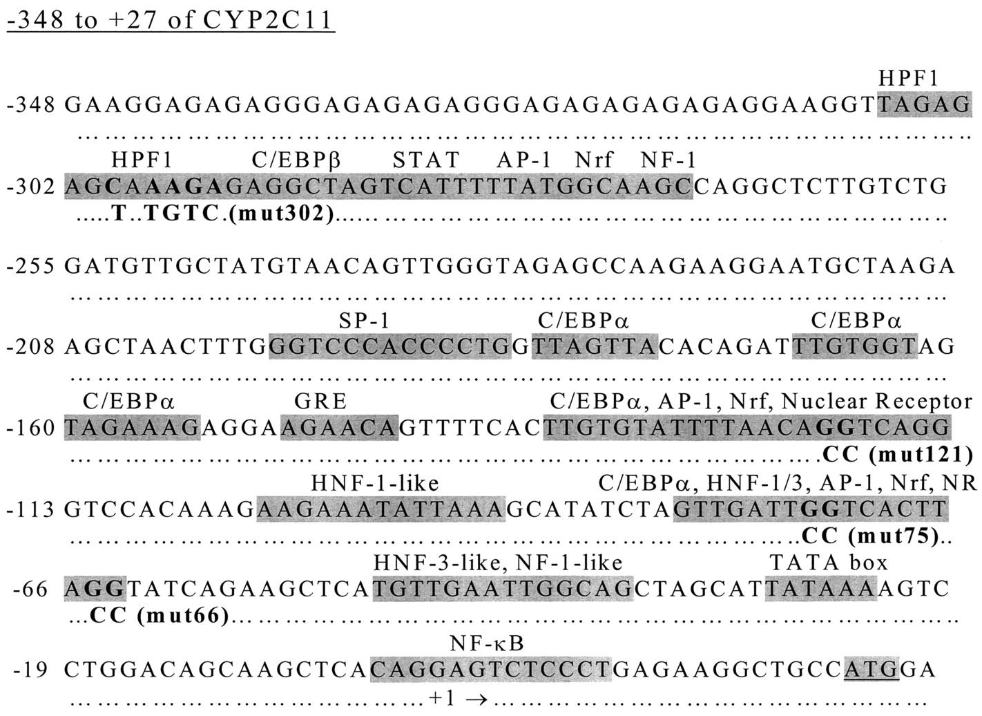
Sequence of -348 to +27 of CYP2C11 5′-flanking sequence. Highlighted sequences indicate transcription factor binding sites as follows: -307 to -287, HPF1 site (Ström et al., 1995); -297 to -270, cluster of putative half-sites for C/EBPβ, signal transducer and activator of transcription, AP-1, Nrf, and NF-1 (MatInspector/TRANSFAC); -197 to -185, putative SP-1 site (MatInspector/TRANSFAC); -183 to -177, -169 to -162, -160 to -154, -135 to -127, -81 to -74, C/EBPα sites (Buggs et al., 1998); -149 to -144, GRE half-site (Ström et al., 1994); -131 to -115, Putative half-sites for AP-1 and Nrf (MatInspector/Transfac); -121 to -116, putative nuclear receptor half-site (MatInspector/Transfac); -103 to -91, -81 to -69, HNF-1–like site (Sundseth et al., 1992); -81 to -64, cluster of putative half-sites for AP-1, Nrf, and estrogen receptor (MatInspector/Transfac); -75 to -65, -50 to -40, HNF-3–like sites (Sundseth et al., 1992); -50 to -37, NF-1–like site (Ström et al., 1994); -29 to -24, TATA box (Morishima et al., 1987); +1, transcription start site (Morishima et al., 1987); -3to +10, nuclear factor-κB site (Iber et al., 2000); +23, translational start site (Morishima et al., 1987). Sequences in boldface type indicate position of mutations and the identities of those mutations are indicated below the wild-type sequences.
The -348 to +27 region of CYP2C11 was also analyzed using MatInspector (ver. 2.2) and TRANSFAC transcription factor database (ver. 4.0) to identify additional putative transcription factors binding sites that might be involved in CYP2C11 regulation (Quandt et al., 1995). The MatInspector/TRANSFAC analysis revealed several clusters of transcription factor half-sites (Fig. 6). Between -297 and -270, there is a cluster of putative half-sites for activator protein-1 (AP-1), signal transducer and activator of transcription, C/EBPβ, Nrf, and NF-1. Between -131 and -115 is another cluster of putative half-sites for AP-1 and Nrf, as well as a perfect nuclear receptor half-site, “AGGTCA” at -121. A third cluster of sites is located between -81 and -64 and contains putative AP-1, Nrf, and estrogen receptor half-sites. A putative SP-1 site was identified at -197 to -185. The functional significance of the sites identified by MatInspector/Transfac is not known.
Effects of Mutations in Putative Transcription Factor Binding Sites on Basal Expression and Negative Regulation of CYP2C11. A series of CYP2C11 constructs containing mutations in putative transcription factor binding sites were generated using overlap extension PCR. The construct “mut302” contains a mutation in the HPF1-site identified by Ström et al. (1995), a site homologous to HNF-4 regulatory sites in rabbit CYP2C genes. Although Ström et al. found that this site did not bind HNF-4, it did bind other factors in nuclear extracts and bind members of the chicken ovalbumin upstream promoter transcription factor family. Mutation of this element resulted in loss of binding of this family but had little effect of transcriptional activity (Ström et al., 1995). In the current study, a mutation strategy identical to that of Ström et al. (1995) was used to produce mut302 (Fig. 6). This mutation had a small effect on basal expression of the 2C11 reporter constructs but no effect on negative regulation by DHEA or nafenopin in either the presence or the absence of PPARα (Fig. 7, A and B).

Mutation of a nuclear receptor half-site at -121 diminishes basal expression of CYP2C11, and mutation of a putative half-site at -75 diminishes negative regulation by DHEA and nafenopin. HepG2 cells were transfected with wild-type 563/2C11-Luc (wt) or 563/2C11-Luc containing mutations as indicated. Cells were cotransfected with pSG5 (A) or pSG5-PPARα (B) and treated 24 h later with 35 μM nafenopin (NAF) or DHEA. Results are expressed as the mean ± S.D. of quadruplicate wells. *, p < 0.05, significantly different from DMSO treatment.
Next, a construct containing a mutation in the perfect nuclear receptor half-site at -121 was generated named “mut121” (Fig. 6). The mutation of the highly conserved GG to CC has previously been shown to diminish PPARα/RXRα binding to DNA (Varanasi et al., 1996). Surprisingly, mutation of this nuclear receptor half-site resulted in greatly diminished basal expression (Fig. 7). The expression of mut121 was 20 to 30% of that of the wild-type construct and was approximately equivalent to that of the lowest expressing deletion construct, 108/2C11-Luc. In addition, mut121 was still negatively regulated by nafenopin and DHEA, suggesting that this site is not involved in negative regulation by peroxisome proliferators but is involved in the basal expression of CYP2C11 in HepG2 cells.
Deletion analysis identified the region between -108 and -60 of CYP2C11 as the negative regulatory region for both DHEA and nafenopin. There is a cluster of putative transcription factor binding sites in this region, including HNF-1- and HNF-3-like sites as well as half-sites for AP-1, Nrf, and an imperfect nuclear receptor half-site. “Mut75” contains a GG-to-CC mutation at -75 bp and “mut66” contains a GG-to-CC mutation at -66 bp relative to the transcription start site (Fig. 6). Mut66 showed a small decrease in basal expression but had no effect on negative regulation. However, mut75 showed a slight decrease in basal expression and complete loss of negative regulation by DHEA and nafenopin. This indicates that the GG at -75 is required for the negative regulation of CYP2C11 by these chemicals. This GG is part of a sequence that may be an imperfect nuclear receptor binding site (TGGTCA). This sequence is also part of the HNF-1- and HNF-3–like sites identified by Sundseth et al. (1992).
PPARα/RXRα Heterodimers Do Not Bind DNA Anywhere in the -348-to- +25 Region of CYP2C11. Results of 5′-deletion and mutation analyses indicated that the peroxisome proliferator-responsive region of CYP2C11 was between -108 and -60 relative to the transcription start site and required the GG nucleotides at -75 bp. To determine whether PPARα/RXRα binds to DNA in this region, gel shift analyses were carried out. In vitro transcribed and translated PPARα and RXRα were incubated with a positive control oligonucleotide containing the PPRE from the rat fatty acyl coA oxidase gene or an oligonucleotide probe containing -86 to -56 of CYP2C11. As expected based on numerous other studies, neither PPARα nor RXRα bound to DNA in the absence of heterodimer partner, but when both PPARα and RXRα were included, binding to the positive control probe was observed (Fig. 8). However, under identical conditions, no binding to the CYP2C11 probe was observed. A second related double-stranded probe containing -91 to -67 of CYP2C11 spanning a 5-bp region upstream of the previous probe was tested; PPARα/RXRα did not bind this oligonucleotide either (data not shown). Additional gel shift experiments were carried out with longer probes containing either -348 to -200 or -200 to +25 of CYP2C11. There was no evidence of PPARα/RXRα binding to CYP2C11 DNA in these experiments either (data not shown). These results suggest that negative regulation of CYP2C11 by PPARα is not mediated by direct binding of the receptor to DNA.

PPARα/RXRα heterodimers bind a consensus PPRE but do not bind DNA sequence in the PPARα-responsive region of CYP2C11. Radiolabeled oligonucleotides containing either the ratty fatty acyl coA oxidase PPRE or -86 to -56 of CYP2C11 were incubated with reticulocyte lyastes expressing PPARα, RXRα, or both, as described under Materials and Methods. DNA-protein complexes were resolved on 4% polyacrylamide gels. A representative PhosphorImage is shown.
Discussion
The purpose of the current work was to examine the mechanism of negative regulation of CYP2C11 by DHEA and to compare the effects of DHEA with classic peroxisome proliferators, such as nafenopin. Initial experiments using cell-based reporter assays indicated that the mechanism for regulation of CYP2C11 by nafenopin required coexpression of PPARα, whereas negative regulation by DHEA did not. Further experiments revealed that the responsive region of CYP2C11 for both chemicals was between -108 and -60 bp relative to the transcription start site. In addition, effects of both DHEA and nafenopin plus PPARα were abolished when a mutation was introduced at -75 bp relative to the CYP2C11 transcription start site. These results suggest that DHEA and nafenopin affect the same DNA binding region with the same ultimate consequences for CYP2C11 expression, but the upstream mechanism leading to the response differs between DHEA and nafenopin.
Previous studies measuring DHEA-induced changes in gene expression used only high doses of DHEA (0.45% DHEA in diet). This dose is known to cause changes in gene expression associated with peroxisome proliferation. However, other studies have demonstrated that DHEA at lower doses (0.012, 0.06, and 0.2% DHEA in diet) are chemoprotective (Lubet et al., 1998). Therefore, we wanted to address whether gene expression changes at high DHEA doses were different that those at lower DHEA doses. The current study shows that this is the case. CYP2C11 mRNA was either not affected or was slightly increased at the lower DHEA doses, but was suppressed only at the highest DHEA doses. We have also noted that induction of CYP3A23 mRNA by DHEA shows a similar bell-shaped dose-response curve, although there is no suppression at the highest DHEA dose (S. L. Ripp and R. A. Prough, unpublished observations). In contrast, induction of CYP4A1 mRNA showed a consistent dose-dependent increase at all DHEA doses. Induction of CYP4A1 by DHEA is known to be a PPARα-dependent process, whereas induction of CYP3A23 is thought to be a pregnane X receptor-dependent process (Ripp et al., 2002). The dose-response data for CYP2C11 regulation by DHEA suggests, but does not prove, that there may be both PPARα-dependent and -independent components of gene regulation, depending on the dose.
To examine the molecular mechanism of negative regulation, reporter assays were developed in several cell lines. All three cell lines tested, HepG2, H4IIE, and CV-1, could support activation of a PPRE-containing reporter gene in the presence of cotransfected PPARα. Activation was enhanced in all three cell lines in the presence of nafenopin. However, DHEA did not activate the PPRE reporter system in any of the cell lines. This is in agreement with the previous studies concluding that DHEA did not activate PPARα in cell-based assays (Peters et al., 1996; Waxman, 1996). When a CYP2C11 reporter gene was used, negative regulation by peroxisome proliferators could be reproduced only in HepG2 cells. Interestingly, similar cell-type specificity was noted for CYP7A1 negative regulation. CYP7A1 was negatively regulated by peroxisome proliferators in HepG2 cells, but not in CV-1 cells (Marrapodi and Chiang, 2000). This indicates a requirement for cellular factors in addition to PPARα to produce negative regulation of these two genes.
Negative regulation of CYP2C11 could be reproduced in HepG2 cells using a luciferase reporter construct containing 1300 bp of CYP2C11 promoter and 5′-flanking region. For the classic peroxisome proliferator nafenopin, coexpression of PPARα was required for negative regulation. Deletion analyses revealed that the responsive region was between -108 and -60 bp relative to the transcription start site. Mutation of a putative nuclear receptor half-site at -75 bp abolished negative regulation, suggesting that this site is required for the transcriptional repression.
There are several potential models to explain how activation of PPARα could result in negative regulation of a gene. Activation of PPARα can result in displacement of other transcription factors from their DNA-binding sites through direct competition for DNA-binding sites. For example, expression of the genes encoding apolipoprotein CIII and transferrin are suppressed by PPARα. Activated PPARα displaces the positive regulatory factor HNF-4 from its binding site, resulting in suppression of expression of these genes (Hertz et al., 1995, 1996). Alternatively, PPARα could directly suppress transcription of a gene through protein-protein interaction with other transcription factors. For example, activation of PPARα results in negative regulation of certain proinflammatory genes by interacting with AP-1 and nuclear factor-κB to disrupt their transactivation abilities (Delerive et al., 1999). Finally, PPARα might indirectly cause repression of gene transcription by modulating the expression of other transcription factors. PPARα activation can increase expression of rev-erbA, which is a negative regulator of other genes (Vu-Dac et al., 1998). PPARα activation can also decrease expression of HNF-4, resulting in decreased expression of HNF-4-regulated genes, as is the case for CYP7A1 (Marrapodi and Chiang, 2000). Because PPARα did not seem to bind to the responsive region of CYP2C11, this would exclude the first model, direct competition with other cellular transcription factors for DNA binding. It is likely that PPARα negatively regulates CYP2C11 by direct protein-protein interaction with other transcription factors or indirectly by modulating expression of other transcription factors. The PPARα responsive region of CYP2C11 contains a cluster of putative transcription factor binding sites, including consensus half-sites for AP-1, ER, and Nrf, as well as HNF-1- and HNF-3–like sites. The negative regulatory effects of PPARα may involve interaction with one or more of these transcription factors.
The effects of DHEA on expression of CYP2C11 in HepG2 cells mapped to the same responsive region as nafenopin, and also was abolished when a mutation was introduced at -75 bp. However, negative regulation by DHEA did not require coexpression of PPARα. There are several potential explanations for these findings. One possibility is that DHEA is acting through a low level of endogenous PPARα present in HepG2 cells. However, this is unlikely, because nafenopin, a more potent activator of PPARα, was able to produce only negative regulation in the presence of coexpressed PPARα. Another possibility is that DHEA treatment induces expression of PPARα in HepG2 cells then follows the same mechanism as nafenopin. This is also unlikely, because DHEA produces additional suppression of CYP2C11 expression even in the presence of coexpressed PPARα. This suggests that DHEA is acting through a mechanism independent of PPARα. DHEA may activate a different member of the nuclear receptor superfamily, which then interferes with basal transcription factors.
We have found that high doses of DHEA can activate pregnane X receptor in vitro (Ripp et al., 2002), and it is likely that high doses of DHEA may activate other nuclear receptors. Recent studies suggest an additional mechanism for the effects of DHEA. DHEA has been suggested to activate membrane receptors, followed by activation of intracellular signaling cascades (Liu and Dillon, 2002). It is known that phosphorylation can alter the transcriptional activity of numerous transcription factors. Therefore, DHEA may be affecting the same downstream target as nafenopin/PPARα; however, DHEA is probably acting through alternative mechanisms to do so.
The exact molecular events leading to negative regulation of CYP2C11 by DHEA and nafenopin remains unclear. However, the work described here identifies a responsive region of DNA and suggests that the mechanism does not involve direct binding of PPARα to this region. This suggests that PPARα is suppressing CYP2C11 expression by modulating expression of or directly interacting with other transcription factors. Future experiments will need to address the basal transcription factors involved in CYP2C11 expression and how peroxisome proliferators affect their function.
Acknowledgments
We are grateful to Eddie Morgan (Emory University, Atlanta, GA) for providing 1300/2C11/CAT and the pGL2-based -344,-148,-112, and -64 2C11 constructs. We also thank Thomas Rushmore for providing PPARα expression vector, Dan Noonan for RXRα expression vector, and Clinton Grubbs (University of Alabama at Birmingham) for providing animal diets containing DHEA.
Footnotes
-
This work was supported by United States Public Health Service grant DK54774 (to R.A.P.) and National Research Service Award F32-ES05927 (to S.L.R.)
-
This work has been previously presented in abstract form [Drug Metab Rev 32(Suppl 2):237, 2000] and related data were published in a symposium minireview (Drug Metab Dispos 29:623–633, 2001).
-
ABBREVIATIONS: DHEA, dehydroepiandrosterone; PPARα, peroxisome proliferator-activated receptor α; RXR, retinoid X receptor; Bp, base pair(s); PCR, polymerase chain reaction; WY-14,643, pirinixic acid [4-chloro-6-(2,3-xylidino)-2-pyrimidinyl)thioacetic acid]; PPRE, peroxisome proliferator responsive element; DHEA-S, dehydroepiandrosterone sulfate; DMSO, dimethyl sulfoxide; CAT, chloramphenicol acetyltransferase; CMV, cytomegalovirus; C/EBP, CCAAT/enhancer-binding protein; HNF, hepatocyte nuclear factor; AP-1, activator protein 1; NF-1, nuclear factor 1
-
↵1 Current address: Pfizer Global Research and Development, Eastern Point Rd., Groton, CT 06340
- Received September 9, 2002.
- Accepted April 2, 2003.
- The American Society for Pharmacology and Experimental Therapeutics


