Advertisement

Main menu

  • Home
  • Articles
    • Current Issue
    • Fast Forward
    • Latest Articles
    • Archive
  • Information
    • Instructions to Authors
    • Submit a Manuscript
    • FAQs
    • For Subscribers
    • Terms & Conditions of Use
    • Permissions
  • Editorial Board
  • Alerts
    • Alerts
    • RSS Feeds
  • Virtual Issues
  • Feedback
  • Other Publications
    • Drug Metabolism and Disposition
    • Journal of Pharmacology and Experimental Therapeutics
    • Molecular Pharmacology
    • Pharmacological Reviews
    • Pharmacology Research & Perspectives
    • ASPET

User menu

  • My alerts
  • Log in
  • My Cart

Search

  • Advanced search
Drug Metabolism & Disposition
  • Other Publications
    • Drug Metabolism and Disposition
    • Journal of Pharmacology and Experimental Therapeutics
    • Molecular Pharmacology
    • Pharmacological Reviews
    • Pharmacology Research & Perspectives
    • ASPET
  • My alerts
  • Log in
  • My Cart
Drug Metabolism & Disposition

Advanced Search

  • Home
  • Articles
    • Current Issue
    • Fast Forward
    • Latest Articles
    • Archive
  • Information
    • Instructions to Authors
    • Submit a Manuscript
    • FAQs
    • For Subscribers
    • Terms & Conditions of Use
    • Permissions
  • Editorial Board
  • Alerts
    • Alerts
    • RSS Feeds
  • Virtual Issues
  • Feedback
  • Visit dmd on Facebook
  • Follow dmd on Twitter
  • Follow ASPET on LinkedIn
Research ArticleSpecial Section – New Models in Drug Metabolism and Transport
Open Access

CRISPR-Cas9: A New Addition to the Drug Metabolism and Disposition Tool Box

M. Karlgren, I. Simoff, M. Keiser, S. Oswald and P. Artursson
Drug Metabolism and Disposition November 2018, 46 (11) 1776-1786; DOI: https://doi.org/10.1124/dmd.118.082842
M. Karlgren
Department of Pharmacy (M.Ka., P.A.), Uppsala University Drug Optimization and Pharmaceutical Profiling Platform, Department of Pharmacy (I.S.), and Science for Life Laboratory (P.A.), Uppsala University, Uppsala, Sweden; and Department of Clinical Pharmacology, Center of Drug Absorption and Transport, University Medicine of Greifswald, Germany (M.Ke., S.O.)
  • Find this author on Google Scholar
  • Find this author on PubMed
  • Search for this author on this site
I. Simoff
Department of Pharmacy (M.Ka., P.A.), Uppsala University Drug Optimization and Pharmaceutical Profiling Platform, Department of Pharmacy (I.S.), and Science for Life Laboratory (P.A.), Uppsala University, Uppsala, Sweden; and Department of Clinical Pharmacology, Center of Drug Absorption and Transport, University Medicine of Greifswald, Germany (M.Ke., S.O.)
  • Find this author on Google Scholar
  • Find this author on PubMed
  • Search for this author on this site
M. Keiser
Department of Pharmacy (M.Ka., P.A.), Uppsala University Drug Optimization and Pharmaceutical Profiling Platform, Department of Pharmacy (I.S.), and Science for Life Laboratory (P.A.), Uppsala University, Uppsala, Sweden; and Department of Clinical Pharmacology, Center of Drug Absorption and Transport, University Medicine of Greifswald, Germany (M.Ke., S.O.)
  • Find this author on Google Scholar
  • Find this author on PubMed
  • Search for this author on this site
S. Oswald
Department of Pharmacy (M.Ka., P.A.), Uppsala University Drug Optimization and Pharmaceutical Profiling Platform, Department of Pharmacy (I.S.), and Science for Life Laboratory (P.A.), Uppsala University, Uppsala, Sweden; and Department of Clinical Pharmacology, Center of Drug Absorption and Transport, University Medicine of Greifswald, Germany (M.Ke., S.O.)
  • Find this author on Google Scholar
  • Find this author on PubMed
  • Search for this author on this site
P. Artursson
Department of Pharmacy (M.Ka., P.A.), Uppsala University Drug Optimization and Pharmaceutical Profiling Platform, Department of Pharmacy (I.S.), and Science for Life Laboratory (P.A.), Uppsala University, Uppsala, Sweden; and Department of Clinical Pharmacology, Center of Drug Absorption and Transport, University Medicine of Greifswald, Germany (M.Ke., S.O.)
  • Find this author on Google Scholar
  • Find this author on PubMed
  • Search for this author on this site
  • Article
  • Figures & Data
  • Info & Metrics
  • eLetters
  • PDF
Loading

Abstract

Clustered regularly interspaced short palindromic repeats (CRISPR)–CRISPR associated protein 9 (Cas9), i.e., CRISPR-Cas9, has been extensively used as a gene-editing technology during recent years. Unlike earlier technologies for gene editing or gene knockdown, such as zinc finger nucleases and RNA interference, CRISPR-Cas9 is comparably easy to use, affordable, and versatile. Recently, CRISPR-Cas9 has been applied in studies of drug absorption, distribution, metabolism, and excretion (ADME) and for ADME model generation. To date, about 50 papers have been published describing in vitro or in vivo CRISPR-Cas9 gene editing of ADME and ADME-related genes. Twenty of these papers describe gene editing of clinically relevant genes, such as ATP-binding cassette drug transporters and cytochrome P450 drug-metabolizing enzymes. With CRISPR-Cas9, the ADME tool box has been substantially expanded. This new technology allows us to develop better and more predictive in vitro and in vivo ADME models and map previously underexplored ADME genes and gene families. In this mini-review, we give an overview of the CRISPR-Cas9 technology and summarize recent applications of CRISPR-Cas9 within the ADME field. We also speculate about future applications of CRISPR-Cas9 in ADME research.

Introduction

Clustered regularly interspaced short palindromic repeats (CRISPR)–CRISPR associated protein 9 (Cas9) is an easily adapted gene-editing technology that can be designed to target most genomic sequences. The major difference between CRISPR-Cas9 and previously favored gene-editing systems—such as the zinc finger nucleases (ZFNs) and the transcription activator–like effector nucleases (TALENs)—is its simplicity. CRISPR-Cas9 DNA targeting only requires the design of a short guide RNA molecule compared with the more complex and expensive protein engineering needed for retargeting ZFNs and TALENs (cf. Gaj et al., 2013; Kim, 2016; Editorial, 2017). The availability and affordability of commercial kits and services means that many biomedical research fields are now using CRISPR-Cas9.

In applied genome editing, researchers have traditionally relied heavily on model organisms such as bacteria, yeast, fruit fly, and rodents. In part, this was because these were the only species for which good tools for genetic manipulation were available. The need for faster gene editing led to the development of custom nucleases like ZFNs and TALENs that induce double strand breaks (DSBs) in a predefined region in the gene of interest (Gaj et al., 2013). Only the ZFN technology has been applied within the field of absorption, distribution, metabolism, and excretion (ADME). There, it has been used for selective knockout of drug transporters in C2BBe1 cells (a subclone of Caco-2, which is a widely used model for investigating transporter-mediated intestinal efflux of drugs) (Pratt et al., 2012; Sampson et al., 2015). Similarly, ZFN has been used to knock out endogenous canine transporters in Madin-Darby canine kidney (MDCK) II cells (Gartzke et al., 2015). It has also been used in HepaRG cells, an alternative to primary human hepatocytes, to knock out the nuclear pregnane X receptor (PXR), a receptor that regulates the expression of drug-metabolizing enzymes and drug transporters (Williamson et al., 2016). Despite these successful studies, neither ZFN nor TALEN have become widespread methods within the field of ADME. One reason may be the expensive and demanding protein engineering needed.

CRISPR-Cas9 has made it possible to edit genes in virtually all organisms for a fraction of the cost compared with alternative technologies. Moreover, CRISPR-Cas9 allows modification of the genes of interest at more physiologically relevant levels (i.e., regulated by an indigenous promoter) rather than by overexpression of the gene of interest. In addition to manipulation of individual genes, CRISPR-Cas9 can even be used to eliminate an entire chromosome (Zuo et al., 2017). This has been demonstrated in human pluripotent stem cells with chromosome 21 trisomy and in cancer cells; highlighting potential therapeutic applications for the CRISPR-Cas9 technology. Furthermore, it is possible to study multiple genes simultaneously, thus creating an intricate network for studying or even curing diseases. One example of such simultaneous gene editing is the knockout of 62 porcine endogenous retroviruses in an immortalized porcine cell line (Yang et al., 2015). In a follow-up study, the same technology was used to generate porcine endogenous retrovirus-free piglets, a strategy that would increase safe and effective organs for pig-to-human xenotranplantations (Niu et al., 2017). However, the in vivo application of CRISPR-Cas9 in humans was impeded after a report in 2015, in which CRISPR-Cas9 gene editing in human embryos showed low rates of homology directed repair (HDR) and apparent off-target effects (i.e., alterations in genes unrelated to the target genes) (Liang et al., 2015). Thus, there are crucial safety issues that have to be solved before broader in vivo applications become a reality. Despite this setback, there are ongoing clinical trials involving CRISPR-Cas9 [https://clinicaltrials.gov/ (accessed on May 20, 2018)]. The first of these focuses on T-cells engineering for treatment of metastatic small cell lung cancer. This study started in 2016 and is expected to be completed during 2018. The engineered T-cells were given to the first patient in October 2016 [https://newatlas.com/crispr-gene-editing-first-human-trial/46453/ (accessed on May 20, 2018)], and the results are not yet published.

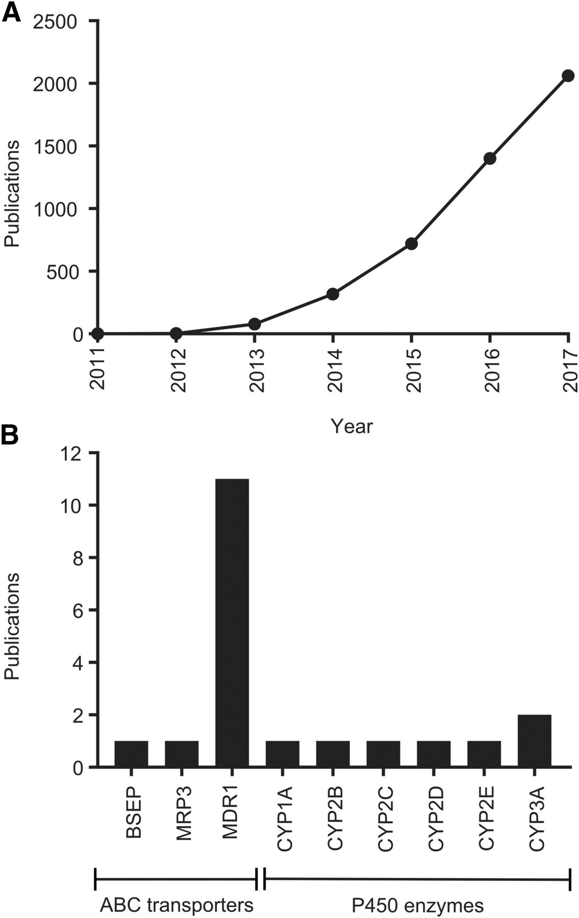
The number of publications regarding CRISPR-Cas9 has increased rapidly (Fig. 1A), from single articles by the originators of the technology in 2011 and 2012, to more than 2000 articles published in 2017. CRISPR-Cas9 is now also being applied within the ADME field (Fig. 1B; Table 1). In this context, it has been mainly used for the generation of in vitro and in vivo knockout models, in parallel with other approaches. Importantly, technologies such as CRISPR-Cas9 also give the opportunity to deorphanize and characterize ADME gene families that are currently underexplored, such as the solute carrier transporter family (César-Razquin et al., 2015). In this mini-review, we give an overview of the application of CRISPR-Cas9 gene editing for ADME studies of drugs and xenobiotics.

Fig. 1.
  • Download figure
  • Open in new tab
  • Download powerpoint
Fig. 1.

(A) Number of publications per year mentioning CRISPR-Cas9. Data were generated by PubMed search using the terms “CRISPR” and “Cas9.” (B) Total number of publications for CRISPR-Cas9 gene editing of ADME genes/proteins. Data were generated by PubMed search using the terms “CRISPR” and “Cas9” and the respective human gene or protein name. All hits were screened and publications were excluded if CRISPR-Cas9 had not been used to edit an ADME gene. If homologous genes from other species had been edited, those publications were included in this compilation. The ADME genes included in the analysis are the solute carrier and ATP-binding cassette (ABC) transporters identified as involved in drug ADME by the International Transporter Consortium (Giacomini et al., 2010) and the most important cytochrome P450 (P450) and uridine diphosphate-glucuronyltransferase enzymes (Evans and Relling, 1999; Williams et al., 2004). The PubMed search was performed May 18, 2018, i.e., articles published after that are not included in this compilation.

View this table:
  • View inline
  • View popup
TABLE 1

Overview of ADME and ADME-related genes edited using CRISPR-Cas9

How Does CRISPR-Cas9 Work?

In nature, CRISPR-Cas9 is an adaptive, acquired immune system, mediated by RNA, which protects eubacteria and archaea from viruses or plasmids (Barrangou et al., 2007). In bacteria, two RNA molecules, together with the endonuclease Cas9, bind to a target sequence. One of the RNA molecules (trans-activating CRISPR RNA) is species conserved and acts as a scaffold that binds Cas9 itself. The other RNA molecule (CRISPR RNA) is variable and confers specificity to a target genome DNA. For gene-editing purposes the technology was simplified and made more efficient by combining both RNAs into a single guide RNA (sgRNA) (Jinek et al., 2012). The sgRNA and Cas9 form a sgRNA-Cas9 complex that binds to its target sequence (Fig. 2, A and B). An additional requirement of the target sequence is the presence of a protospacer adjacent motif (PAM) (Mojica et al., 2009). If the sgRNA hybridize to DNA immediately 5′ to an adjacent PAM domain the Cas9 endonuclease will create a DSB (Fig. 2B). This DSB can be repaired by the mechanism of nonhomologous end joining (NHEJ) (Overballe-Petersen et al., 2013). This, in turn, may result in small deletions or insertions (indels) that disrupt the targeted gene, resulting in loss of its function (Fig. 2C). Alternatively, if there is a donor DNA template to the targeted region the DSB can be repaired by HDR, thereby allowing for precise replacement of nucleotide(s), introduction of mutations, or insertion of sequences in the targeted region (Gong et al., 2005) (Fig. 2C). The frequency of NHEJ indel formation or successful HDR varies, depending on the model cell line/organism and methods used; however, in general HDR is a rarer event than NHEJ indel formation (Mali et al., 2013). For an overview of CRISPR discovery and history, see Lander (2016).

Fig. 2.
  • Download figure
  • Open in new tab
  • Download powerpoint
Fig. 2.

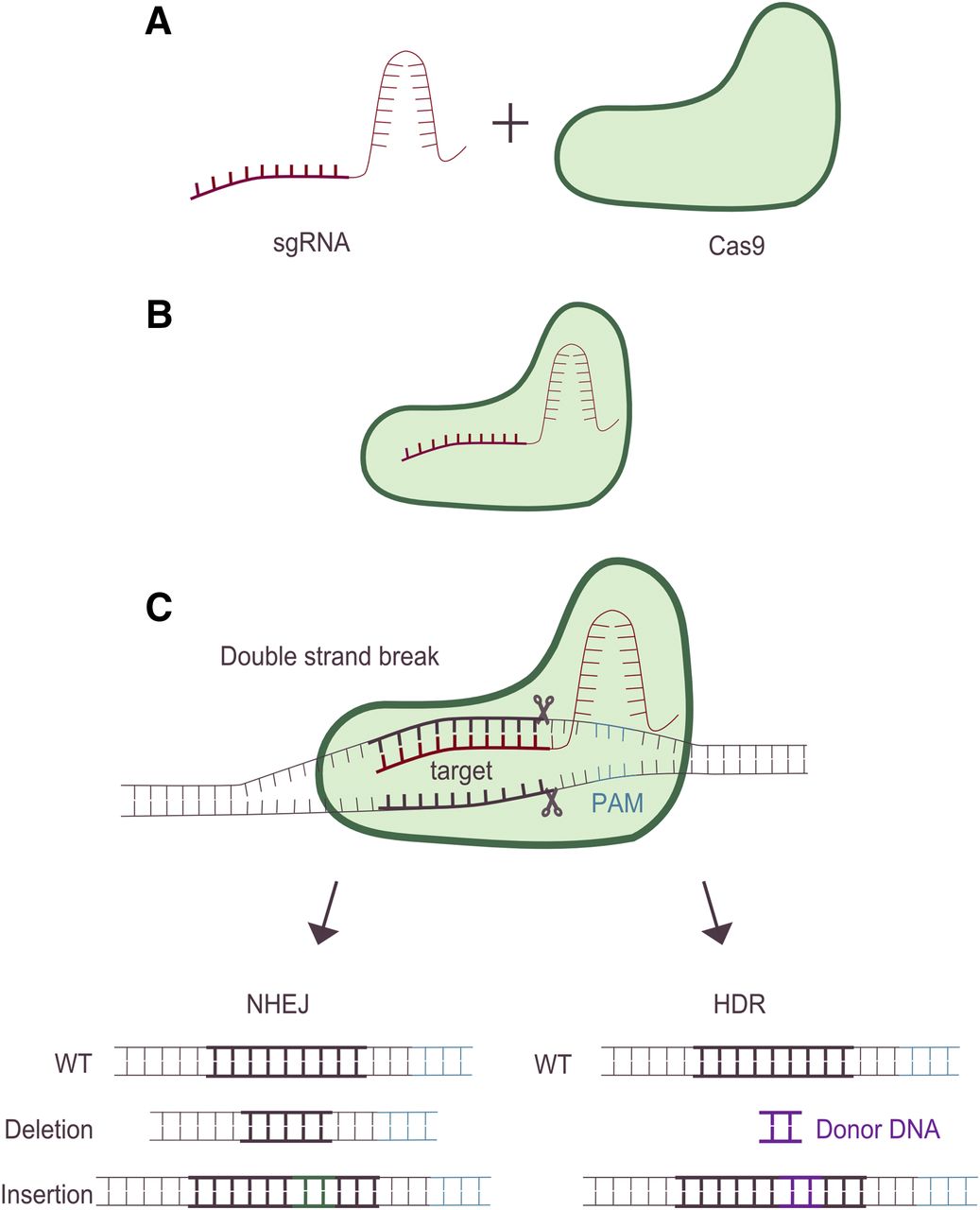
Overview of the CRISPR-Cas9 gene-editing process. For CRISPR-Cas9 gene editing, a sgRNA is designed. This sgRNA consists of two parts, one that is specific for the desired target DNA and one that will interact with the second component in the CRISPR-Cas9 system, the Cas9 endonuclease (A). An important requirement of the target DNA is the presence of a PAM. After formation of a sgRNA-Cas9 complex (B), the complex will bind to the target DNA, and if an adjacent PAM is present Cas9 will cause a DNA DSB three base pairs upstream of the PAM sequence (C). Thereafter, the DSB can be repaired using two different mechanisms, NHEJ or HDR. NHEJ is an error-prone mechanism that will often result in small deletions or insertions (indels) that can lead to a disruption in the targeted locus. Alternatively, if a homology-containing donor-DNA template to the targeted region is present, the DSB can be repaired instead by HDR, thereby allowing for precise replacement, introduction of mutations or insertion of sequences in the targeted region. WT denotes wild type.

The CRISPR-Cas9 system primarily used for gene editing comes from Streptococcus pyogenes. However, Cas9 proteins from other organisms are being explored to increase the flexibility and expansion of application areas (as discussed subsequently). CRISPR-Cas9 has developed from in-house design to predesigned sgRNAs and premade CRISPR kits available from multiple vendors. In addition, software is available, e.g., for sgRNA design, analysis of off-target effects, etc. [e.g., http://crispr.mit.edu/, http://www.e-crisp.org/E-CRISP/, https://crispr.dbcls.jp/, http://www.rgenome.net/cas-offinder/, and http://www.biootools.com/ (accessed on May 23, 2018)]. The commercial availability of kits and software extends the possibilities of the CRISPR-Cas9 technology to researchers within many fields.

Improved CRISPR-Cas9 Technologies.

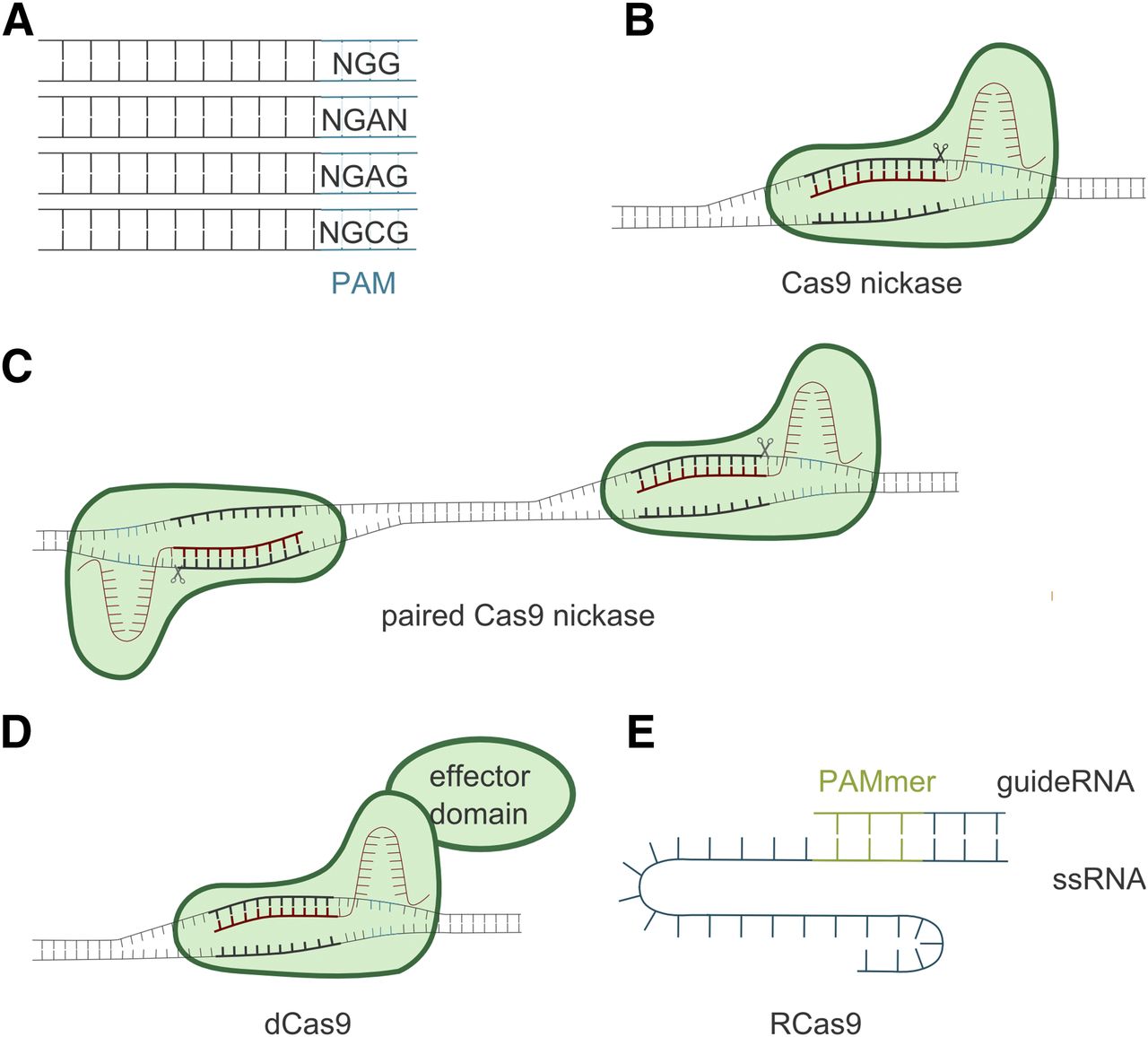
New or improved CRISPR technologies are constantly being developed, offering more opportunities and diverse applications (Sander and Joung, 2014). One limitation of CRISPR-Cas9 is set by the PAM motif—sites must contain the NGG PAM sequence for gene editing to occur (see Fig. 2C). To overcome this limitation, other species, endonucleases, and PAM recognition sequences are being explored, generating even more PAM motif variants (Fig. 3A) (see Nakade et al. (2017), and references therein). Together, these expand the precision and applicability of CRISPR as a gene-editing tool. Recently, a thermostable Cas9 was described with activity up to 70°C. This endonuclease exhibited significant stability in diluted human plasma, potentially providing a new opportunity for therapeutic applications via intravenous administration (Harrington et al., 2017).

Fig. 3.
  • Download figure
  • Open in new tab
  • Download powerpoint
Fig. 3.

The CRISPR-Cas9 technology is constantly being expanded to improve specificity and increase its versatility. Such improvements include more flexible PAM requirements by modification of the Cas9 endonuclease from Stretococcus pyogenes, or by use of endonucleases from other species (A). Another improvement is the engineering of Cas9 into a Cas9 nickase (Cas9D10A) that only cuts one of the DNA strands. This nickase can be used both in single (B) or paired (C) strands, thereby reducing the risk of indels at off-target sites. Another engineered Cas9 is dCas9, which does not cut the DNA, but can be coupled to an effector domain, like a transcriptional activator/repressor, epigenetic modifiers, or fluorescent proteins (D). The Cas9 endonuclease has also been modified to target single-stranded RNA (ssRNA) (RNA-targeting Cas9 or RCas9) instead of double-stranded DNA (E).

One approach to overcome off-target effects is to use mutated Cas9 (Cas9D10A) that introduce a single-strand break (a nick) in the target DNA instead of a DSB (Fig. 3B) (Cong et al., 2013). Hence, two sgRNAs (one on the sense and one on the antisense strand) are needed to produce a DSB and give rise to a 5′ overhang (Fig. 3C). This will minimize the off-target effects, whereas the on-target effects will occur at the same rates as with wild-type Cas9 (Ran et al., 2013).

A modified Cas9, called nuclease deficient Cas9 (dCas9), can be applied for other technical improvements (Gilbert et al., 2013). The dCas9 protein lacks endonuclease function, but it can be further modified by coupling it with a functional protein domain with different activity, e.g., a transcriptional repressor (CRISPR interference) or activator (CRISPR activation) (Lo and Qi, 2017). With the ability of sgRNA to bind to a specific target sequence, the coupled dCas9 can be targeted to the specific gene of interest (Fig. 3D).

Similarly, the dCas9 protein can be coupled to epigenetic modifiers for locus-specific alteration of acetylation or methylation patterns and subsequent suppression/induction of gene expression (cf. Lo and Qi, 2017; Murovec et al., 2017). Other applications of dCas9 include fusion to fluorescent proteins for visualization of specific DNA sequences in living cells (Chen et al., 2013), or fusion to the unspecific endonuclease domain of FokI. Since FokI is only active as a dimer, combining the fused dCas9-FokI with two specific and adjacent sgRNAs increases the specificity of cleavage and reduces the risk for off-target effects (Guilinger et al., 2014).

Cas9 protein has also been modified to target single-stranded RNA (O’Connell et al., 2014). This modified Cas9 is referred to as RNA-targeting Cas9. In this approach, the PAM sequence is mismatched compared with the DNA sequence that allows RNA-exclusive targeting. It is supplied as part of the oligonucleotide that will hybridize to the target RNA (Fig. 3E). Thus far, RNA-targeting Cas9 has been used, e.g., for RNA cleavage, isolation, and tracking in living cells (O’Connell et al., 2014; Nelles et al., 2016). Similarly, a new endonuclease, Cas13, is being used to detect single molecules of RNA or DNA (Shmakov et al., 2017). This endonuclease has been applied in diagnostics to detect single-stranded RNA of Zika and Dengue viruses, as well as mutations in patient liquid samples (Gootenberg et al., 2018).

CRISPR-Cas9 for Generating ADME Models

CRISPR-Cas9, like RNA interference, has been used as a tool for target identification in drug discovery (Taylor and Woodcock, 2015; Xue et al., 2016). Since CRISPR-Cas9 is a relatively new technology, the number of CRISPR-Cas9 studies targeting ADME genes is limited. Nevertheless, by 2013 the first reports of CRISPR-Cas9 gene editing of ATP-binding cassette transporters were presented. In these cases, the gene edited was the CFTR gene (ABCC7) (Schwank et al., 2013) and the white gene (belonging to the ABCG family) regulating eye pigmentation in fruit fly (Kondo and Ueda, 2013), i.e., transporters not considered to be ADME proteins. Two years later, in 2015, the first articles about gene editing of solute carrier transporters were published. These articles described the generation of SLC31A1 and SLC31A2 knockouts, two copper transporters that are also involved in cisplatin and carboplatin transport (Tsai et al., 2015). A year later, in 2016, the first mammalian in vivo knockout ADME models were developed in Sprague-Dawley or Wistar rats. Both models focused on drug-metabolizing cytochrome P450 (P450) enzymes and the genes knocked out were Cyp2e1 (Wang et al., 2016) or the Cyp2d gene cluster (Yoshimi et al., 2016). In the latter publication, a humanized rat model was also generated since the human CYP2D6 gene was knocked in instead of the rodent Cyp2d genes. Since then, CRISPR-Cas9 has been applied in a number of studies focusing on drug-metabolizing enzymes, drug transporters, nuclear receptors, or elements involved in the regulation of ADME genes (for an overview, see Table 1).

Drug-Metabolizing Enzymes

In Vitro.

To our knowledge, only one in vitro P450 model has been established using CRISPR-Cas9. It was based on the human hepatocyte cell line Huh-7 (Dorr et al., 2017). Here, CRISPR-Cas9 editing of the CYP3A5 locus established heterozygous and homozygous in vitro models for the CYP3A5*1 allele. The Huh-7 cell line is homozygous for the CYP3A5*3 allele, an allele with a splice defect resulting in an inactive enzyme, which is also the most prevalent allele in Caucasian populations (Zhou et al., 2017). However, the CYP3A5*1 allele has an allele frequency of up to 56% in non-Caucasian populations, necessitating in vitro models that reflect other genotypes. The gene editing of CYP3A5 was successful and resulted in elevated CYP3A5 mRNA levels and increased metabolism of the two CYP3A5 substrates midazolam and tacrolimus (Dorr et al., 2017).

In Vivo.

Prior to the CRISPR-Cas9 technology, in vivo knockout P450 models, humanized P450 models (where animals express the human P450 ortholog), or chimeric models (where human hepatocytes were transplanted and allowed to repopulate the host liver) were used to study ADME- and toxicity-related problems (Tateno et al., 2004; Cheung and Gonzalez, 2008; Hasegawa et al., 2011; Jiang and Yu, 2012; Kazuki et al., 2013; Scheer et al., 2013). However, the major applications of these models involve, e.g., studies of developmental or physiologic functions, disease mechanisms, validation of target genes, identification of off-targeted effects for drugs and drug candidates, etc.

A number of in vivo CRISPR-Cas9 knockout models have been established for P450 enzymes, including five articles on the generation of mammalian knockout and/or knockin models. These models that are based on Wistar or Sprague-Dawley rats and/or C57BL/6 mice targeted genes coding for enzymes in drug-metabolizing P450 families 2 and 3. In the first model the rat Cyp2d gene locus (containing Cyp2d1-5) was knocked out in Wistar rats and replaced with human CYP2D6 (Yoshimi et al., 2016). Thus, this generated both a Cyp2d knockout rat and a humanized CYP2D6 rat. Unfortunately, no functional characterization was done and to our knowledge no follow-up studies with these in vivo models have been published. In other models Cyp2e1 (Wang et al., 2016) or Cyp2c11 (Wei et al., 2018) were knocked out in Sprague-Dawley rats. The CRISPR-Cas9 technology has also been used to generate a double-knockout rat model in which Cyp3a1 and Cyp3a2 are knocked out (Lu et al., 2017). The Sprague-Dawley knockout models were generated to establish in vivo models for investigating physiologic functions and xenobiotic metabolism by the specific enzyme(s) of interest. In vitro (using liver microsomes from the knockout rats) and in vivo studies using prototypical P450 substrates showed that the metabolic function of the targeted enzyme(s) was indeed impaired in all knockout models. However, all models showed compensatory upregulation or downregulation of other P450s involved in drug metabolism. Moreover, these models also showed other differences, e.g., altered serum concentrations of testosterone (Lu et al., 2017) or alkaline phosphate (Wang et al., 2016) and reduced fertility (Wei et al., 2018). Some of these differences can be attributed to the function of the knocked out gene, e.g., CYP3A and testosterone metabolism, whereas others are unexpected. This suggests that such physiologic alterations need to be taken into consideration when comparing ADME data from the knockout models with data from the wild type.

Besides the rat models, a C57BL/6 mouse model was established, with three Cyp2b genes—Cyp2b9, Cyp2b10 and Cyp2b13—knocked out (Kumar et al., 2017). Interestingly, there were few compensatory changes in expression levels of other P450 enzymes in the triple knockout model. The significant changes that were observed were in female mice for which mRNA expression of Cyp2a4, Cyp2c40, and Cyp3a13 were downregulated.

Drug Transporters

In Vitro.

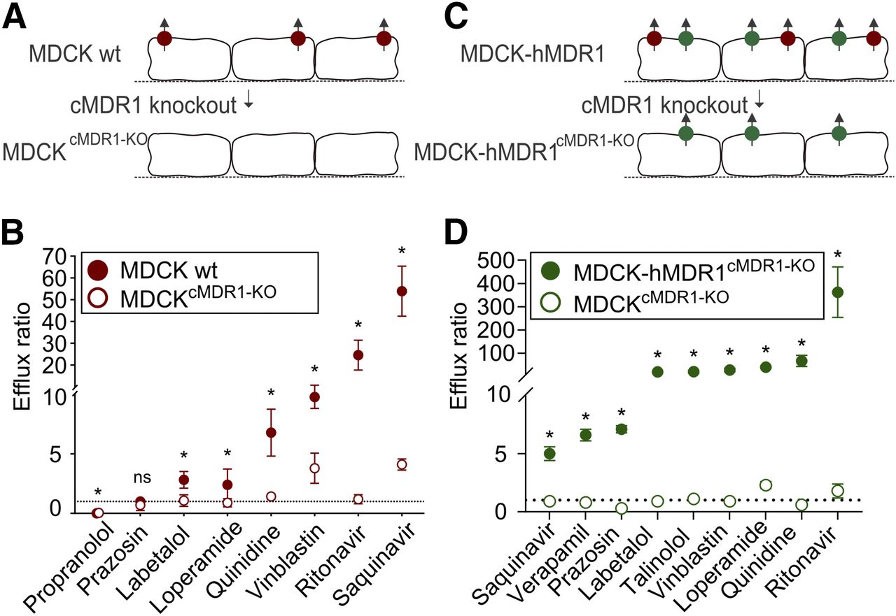
CRISPR-Cas9 gene editing in cell culture models for drug transport has mainly focused on ABCB1/MDR1 (Fig. 1B). To the best of our knowledge, the first paper regarding MDR1 and ADME was published by us in 2016 (Simoff et al., 2016) with a follow up 1 year later (Karlgren et al., 2017). The aim in both studies was to knock out the endogenous canine ABCB1/MDR1 expression in two MDCK cell lines—the wild-type cell line and the cell line already overexpressing the human MDR1 transporter. The monolayer-forming MDCK cell line has been used for decades for heterologous expression of single or multiple drug transporters and subsequent transport studies, and it is favored as a predictive drug efflux model. Unfortunately, due to a large substrate overlap the endogenous background of canine MDR1 interferes with the overexpressed human ABCB1/MDR1 (Kuteykin-Teplyakov et al., 2010; Wang et al., 2013; Karlgren et al., 2017). This makes it difficult to interpret and derive correct kinetic variables for the human homolog. We reasoned that selective knockout of canine ABCB1 would provide an MDCK transport model without interference from the endogenous background. After transfection of the wild-type and overexpressing MDCK cells with plasmids containing sgRNA-Cas9 and clonal selection, two isogenic cell lines were generated with identically disrupted canine ABCB1 genes. Sequencing analysis and liquid chromatography–tandem mass spectrometry–based targeted proteomics verified that the clones were indeed completely lacking canine MDR1 expression. The disruption was homozygous and included four nucleotide deletions that produced frame-shift and premature stop codons in both cell lines. Furthermore, functional analysis showed no efflux of prototypical MDR1 substrates in the knockout (KO) cell line based on wild-type MDCK (MDCKcMDR1-KO) (Fig. 4, A and B). In comparison, the efflux ratios in the original MDCK wild-type cells were significantly higher, indicating that MDCKcMDR1-KO indeed was completely lacking canine MDR1 (Fig. 4B).

Fig. 4.
  • Download figure
  • Open in new tab
  • Download powerpoint
Fig. 4.

Overview of MDCK cell models where the canine ABCB1/MDR1 has been knocked out using CRISPR-Cas9. (A) Canine MDR1 (cMDR1) was knocked out in an MDCK wild-type cell line; after clonal selection, a knockout clone was obtained that completely lacks endogenous cMDR1 expression (MDCKcMDR1-KO). (B) Functional validation using Transwell studies and prototypical MDR1 substrates showed significantly lower efflux ratios for MDCKcMDR1-KO compared with the parental cell line and with most efflux ratios being close to unity (dotted line). (C) cMDR1 was knocked out in an MDCK cell line already overexpressing the human MDR1 transporter (hMDR1). Here, a clone with identical gene editing as MDCKcMDR1-KO and completely lacking cMDR1 expression could be identified (MDCK-hMDR1cMDR1-KO). (D) Functional validation using Transwell studies and prototypical MDR1 substrates showed significantly higher efflux ratios compared with MDCKcMDR1-KO. Furthermore, the hMDR1 activity was retained, indicating no editing of the human ABCB1. Data are presented as mean ± standard deviation for one representative experiment performed in triplicate. ns, not significant, *p < 0.05; using student’s t-test. Graphs (B and D) have been previously published in Simoff et al. (2016) and Karlgren et al. (2017), respectively. These graphs were adapted and republished with kind permission from the publisher.

A knockout cell line, MDCK-hMDR1cMDR1-KO, was also developed for the corresponding MDCK cells overexpressing human MDR1. Significant differences in efflux ratios were seen between MDCKcMDR1-KO and MDCK-hMDR1cMDR1-KO, indicating that the homologous human ABCB1 was largely unaffected by the CRISPR-Cas9 knockout process (Fig. 4, C and D). Recently, we used the MDCKcMDR1-KO cell line, which lacks endogenous canine ABCG2/BCRP activity, for heterologous expression of the human drug transporter BCRP (M. Karlgren et al., manuscript in preparation). Since BCRP and MDR1 share many substrates, the absence of interfering canine MDR1 background is an advantage of this novel model.

Five additional articles published between 2016 and 2018 applied a similar approach to knockout ABCB1/MDR1in various cell lines. These investigated if the knockout of ABCB1 could reverse resistance in human cancer derived cell lines (Ha et al., 2016; Liu et al., 2016; Yang et al., 2016b; Takahashi et al., 2017; Norouzi-Barough et al., 2018). As expected, these studies showed decreased ABCB1/MDR1 mRNA or protein expression levels and increased intracellular accumulation, or increased sensitivity in the MDR1 knockout cells using MDR1 substrates such as rhodamine 123, doxorubicin, and/or carfilzomib. Besides this clinically relevant drug transporter (Giacomini et al., 2010), CRISPR-Cas9 gene editing has also been used in editing of a number of other transporters (see Table 1).

In Vivo.

A number of in vivo transporter models for ADME studies were developed before CRISPR-Cas9. Most of these are in vivo transporter knockout models for studies of drug transport. These models allowed studies of the role of a specific transporter in the presence of all other mechanisms influencing drug disposition in vivo, such as drug uptake, distribution, metabolism, and efflux. For overviews of in vivo knockout transporter models, see Jiang and Yu (2012) and Tang et al. (2013). Prominent examples of the applicability of single and multiple in vivo knockout models for studies of transport mechanisms include ABCB1/MDR1 in the blood-brain barrier (Schinkel et al., 1995) and SLCOs/OATPs in Rotor-type hyperbilirubinemia (van de Steeg et al., 2012), respectively. However, a complicating factor is that knockout of a single transporter protein or several transporter proteins can lead to increased expression of other transporter proteins (e.g., Kuroda et al., 2004; Vlaming et al., 2009). Thus far, in vivo CRISPR-Cas9 gene editing of drug transporters has focused on the ATP-binding cassette transporter family (see Table 1), where it has been used to knock out ABCB1/MDR1 homologs in insects (Denecke et al., 2017; Zuo et al., 2018). In both studies the knockout of ABCB1/MDR1 homologs increased the susceptibility to various insecticides. The ABCB1 homolog was also knocked out in zebrafish. The mutated zebrafish embryos showed a reduced efflux of model substrates and also increased accumulation of cadmium chloride and benzo[a]pyrene (Tian et al., 2017). In another study, abcb11b, the ortholog of human ABCB11/BSEP, was knocked out in zebrafish (Ellis et al., 2018). Like humans with impaired BSEP function, the knockout of abcb11b in zebrafish was associated with, e.g., activation of impaired bile salt excretion and autophagy. See Table 1 for a summary of CRISPR-Cas9 in vivo editing of additional transporters.

Regulation of ADME Genes

The CRISPR-Cas9 technology has been applied to study the regulation of ADME genes. An example is the regulation of the drug transporter ABCC3/MRP3 in the human keratinocyte cell line HaCaT (Takechi et al., 2018). In this study, the authors identified a CpG island, i.e., an area rich in CpG dinucleotides often found near or within the promoter or first exon of a gene, and which may be involved in the epigenetic regulation of genes. This island is located approximately 10 kb upstream of the ABCC3 gene and its methylation status correlates negatively with ABCC3/MRP3 mRNA expression levels in human skin samples. The CRISPR-Cas9-induced deletions in this CpG area reduced ABCC3/MRP3 mRNA expression levels in the HaCaT cell line. This study highlights the possibility of using CRISPR-Cas9 for editing/deletion of nontranscribed DNA targets.

CRISPR-Cas9 has been used for studying expression and function of nuclear receptors involved in P450 regulation (see Table 1). The aryl hydrocarbon receptor is involved in induction of, e.g., CYP1A1. To generate model systems for the study of aryl hydrocarbon receptor signaling and its modulation by xenobiotics, a Luciferase reporter cassette was knocked in at the CYP1A1 transcription start site in induced pluripotent stem cells (iPSCs) using CRISPR-Cas9 (Smith et al., 2016). The authors could monitor aryl hydrocarbon receptor activity by following CYP1A1 expression in these cells. In another study, in human iPSC, CRISPR-Cas9 has been used to tag PXR with the fluorescent protein mCherry (Kim et al., 2018). PXR is involved in the induction of several drug-metabolizing enzymes and transporters (Kandel et al., 2016), but its expression is low in iPSC-derived hepatocytes (Kim et al., 2018). Consequently, this results in low enzyme/transporter levels. The authors reason that the established PXR-mCherry iPSC cell line forms a platform for discovering factors that can increase PXR expression levels during differentiation; however, to date, no such studies have been published. These two articles point to the possibility of following endogenous gene expression with the aid of CRISPR-Cas9. It can be speculated that once established this approach will offer an alternative to other mRNA or protein quantification technologies.

Limitations of CRISPR-Cas9

The CRISPR-Cas9 technology also has its limitations. As mentioned previously, CRISPR-Cas9 gene editing shows apparent off-target effects as well as low efficiency (Liang et al., 2015). DSBs can occur even if a few mismatches exist between the sgRNA and the DNA target. This can result in alterations in genes unrelated to the target gene (Fu et al., 2013). Gene editing at sites with as many as five mismatches has been reported; the rate of such off-target effects can be similar to that of the gene editing at the target site (Fu et al., 2013; Lin et al., 2014).

Potential off-target effects are today often investigated using in silico analysis to avoid target sites with closely resembling sequences. If this is not possible, such off-target sites are investigated for unwanted mutations using polymerase chain reaction and/or DNA sequencing. Nevertheless, it should be noted that a recent report showed that CRISPR-Cas9 editing could also cause single-nucleotide variants in unpredictable off-target sites in vivo in mice (Schaefer et al., 2017). However, that report was retracted during the compilation of this paper since proper genetic background analysis had not been performed (Schaefer et al., 2018). Even so, this shows that—for comprehensive off-target analysis—whole genome sequencing might be the method of choice.

The low efficiency in CRISPR-Cas9 editing is a limitation, especially for in vivo applications. However, even in in vitro studies, e.g., the development of CRISPR-Cas9 knockout models by the NHEJ mechanism, the low efficiency results in the need for extensive screening to find homozygous knockout clones. In our experience, screening of clones enriched using, e.g., green fluorescent protein or antibiotic selection, can result in up to 28% of the screened clones having homozygous deletions (Simoff et al., 2016; Karlgren et al., 2017). This is in line also with other publications reporting similar efficiencies for CRISPR-Cas9 and NHEJ (Moore, 2015; Kosicki et al., 2017), but lower efficiencies for HDR (Mali et al., 2013; Shao et al., 2014). It should be noted that since the gene editing is sequence specific large variation in efficiency is seen for different targets and delivery methods (Kosicki et al., 2017).

The CRISPR-Cas9 technology is also limited by the requirement of a PAM motif in close proximity to the target site. Although endonucleases recognizing alternative PAM sequences are now available (as previously mentioned), this might still constitute a problem when targeting ADME genes that have high sequence identity with other gene family members. In such cases, genomic screening and validation (mRNA/protein expression or activity) might also be more difficult.

After submission of this article, results were published indicating additional problems with CRISPR-Cas9, in particular for its clinical applicability. Haapaniemi et al. (2018) showed that DSBs introduced by CRISPR-Cas9 triggered p53-dependent cell cycle arrest and that this response was independent of the targeted locus. Similarly, Ihry et al. (2018) showed that CRISPR-Cas9-induced DSBs generated a p53-dependent toxic response in human pluripotent stem cells. The targeting of a single locus was sufficient to kill most of the cells (Ihry et al., 2018). This suggests that p53-deficient cells have a growth advantage after CRISPR-Cas9 gene editing. Therefore, if CRISPR-Cas9 is to be used for therapeutic purposes, p53 function needs to be assured to avoid the risk of cancer. Another serious problem, raised by Kosicki et al. (2018), is that CRISPR-Cas9 gene editing may result in large deletions and complex gene rearrangement at the target site. Such alterations are undetectable by the short-range polymerase chain reaction assays commonly used to verify gene modifications. Lesions and crossover events were also detected outside of the targeted locus (Kosicki et al., 2018). Together, these findings highlight the importance of comprehensive genetic analysis to reveal and control unexpected gene modifications prior to a more general clinical use of CRISPR-Cas9.

Future Applications for CRISPR-Cas9 within the ADME Field

Despite the restrictions mentioned, the simplicity, programmability, versatility, and comparably low costs of CRISP-Cas9, together with the rapid methodological development and improvement, indicate a huge potential for a variety of applications in the ADME field. At this point in time, CRISPR-Cas9 has mainly been used to generate in vitro or in vivo knockout models of ADME genes. One major advantage with CRISPR-Cas9 knockout models is the complete elimination of background expression, which will facilitate investigations of clinically relevant drug transporters and drug-metabolizing enzymes. CRISPR-Cas9 knockout models will also be useful in studies of putative ADME genes, such as orphan solute carrier family members (César-Razquin et al., 2015; Lin et al., 2015) and other ADME genes of relevance in drug disposition and drug interactions, as drug targets, and that are associated with human disease. With CRISPR-Cas9, one has the opportunity to not just knock out the gene of interest, but also to study the role of individual nucleotides or amino acids as structural/functional constituents, or to humanize animal ADME genes under a native promoter.

An intriguing opportunity with CRISPR-Cas9 is that noncoding DNA can be targeted. Interindividual variation (expression levels, drug-drug interactions, and pharmacokinetics) of ADME proteins can be due to genetic variation in coding regions or promoter regions, but much of the variation is still unexplained. We know that the DNA encoding proteins, which are fairly well studied, represent only a minor part of the human genome. The regulatory role of noncoding DNA is mainly unexplored. Furthermore, genome-wide association studies have shown that most of the associated sites in human diseases are located in these noncoding DNA regions (Wright and Sanjana, 2016). Similar observations have been reported also for ADME proteins, e.g., regarding variation in ATP-binding cassette transporter expression levels (Matsson et al., 2012). Therefore, Wright and Sanjana (2016) proposed that a future application of CRISPR-Cas9 could be to shed light on this intriguing issue.

While many drugs and drug metabolites interact with both drug transporters and drug-metabolizing enzymes in vivo, transport and metabolism are often studied separately in vitro using recombinant proteins, cell lines overexpressing transporters, or drug-metabolizing enzymes. This is because the contribution of an individual transporter/enzyme can be difficult to determine in more complex in vitro models, such as human hepatocytes. As a consequence, we and others have previously developed in vitro models overexpressing different combinations of transporters and enzymes, in which the contribution of an individual transporter/enzyme can more easily be determined (Crespi et al., 2000; Fahrmayr et al., 2012, 2013; Kwatra et al., 2012; Neve et al., 2013). These models are useful but have drawbacks, including endogenous background expression and time-consuming experimental procedures. With CRISPR-Cas9 it is possible to both completely knock out and knock in genes; hence, with this technology we will be able to develop better and easier-to-use models than the traditional ones. For example, transport-metabolism interplay or drug metabolite interactions could be studied under more controlled conditions using such models.

The use of dCas9 for genome docking and the coupling of dCas9 to an effector domain could provide additional opportunities to generate in vitro models that are more predictive of the in vivo situation. Through dCas9 docking/coupling it could become possible (instead of knocking in or knocking out genes) to modulate expression levels of one or several endogenous genes simultaneously, thereby providing models with more in vivo–like expression profiles.

In summary, we have demonstrated the versatility and applicability of the CRISPR-Cas9 technology. By using examples from more basic research areas, our goal was to stimulate increased use of CRISPR-Cas9 in the ADME field. We predict that the advantages of CRISPR-Cas9 will result in a variety of more advanced and better interpretable in vitro and in vivo models for drug metabolism and disposition.

Acknowledgments

We are grateful to Dr. Janneke Keemink for producing the figures included in this paper.

Authorship Contributions

Wrote or contributed to the writing of the manuscript: Karlgren, Simoff, Keiser, Oswald, Artursson.

Footnotes

    • Received June 1, 2018.
    • Accepted August 3, 2018.
  • This work is supported by The Swedish Fund for Research without Animal Experiment (Approval Number 0004) to M.Ka.; the Magnus Bergvall Foundation (Approval No. 2305) to M.Ka.; and the Swedish Research Council (Approval No. 1951) to P.A.

  • https://doi.org/10.1124/dmd.118.082842.

Abbreviations

ADME
absorption, distribution, metabolism, and excretion
Cas9
clustered regularly interspaced short palindromic repeats associated protein 9
CRISPR
clustered regularly interspaced short palindromic repeats
dCas9
nuclease deficient clustered regularly interspaced short palindromic repeats associated protein 9
DSB
double strand break
HDR
homology directed repair
iPSC
induced pluripotent stem cell
KO
knockout
MDCK
Madin-Darby canine kidney
NHEJ
nonhomologous end joining
P450
cytochrome P450
PAM
protospacer adjacent motif
PXR
pregnane X receptor
sgRNA
single guide RNA
TALEN
transcription activator-like effector nuclease
ZFN
zinc finger nuclease
  • Copyright © 2018 by The Author(s)

This is an open access article distributed under the CC BY-NC Attribution 4.0 International license.

References

    1. Aluru N,
    2. Karchner SI,
    3. Franks DG,
    4. Nacci D,
    5. Champlin D, and
    6. Hahn ME
    (2015) Targeted mutagenesis of aryl hydrocarbon receptor 2a and 2b genes in Atlantic killifish (Fundulus heteroclitus). Aquat Toxicol 158:192–201.
    OpenUrlCrossRefPubMed
    1. Angelos MG,
    2. Ruh PN,
    3. Webber BR,
    4. Blum RH,
    5. Ryan CD,
    6. Bendzick L,
    7. Shim S,
    8. Yingst AM,
    9. Tufa DM,
    10. Verneris MR, et al.
    (2017) Aryl hydrocarbon receptor inhibition promotes hematolymphoid development from human pluripotent stem cells. Blood 129:3428–3439.
    OpenUrlAbstract/FREE Full Text
  1. ↵
    1. Barrangou R,
    2. Fremaux C,
    3. Deveau H,
    4. Richards M,
    5. Boyaval P,
    6. Moineau S,
    7. Romero DA, and
    8. Horvath P
    (2007) CRISPR provides acquired resistance against viruses in prokaryotes. Science 315:1709–1712.
    OpenUrlAbstract/FREE Full Text
    1. Bellec J,
    2. Bacchetta M,
    3. Losa D,
    4. Anegon I,
    5. Chanson M, and
    6. Nguyen TH
    (2015) CFTR inactivation by lentiviral vector-mediated RNA interference and CRISPR-Cas9 genome editing in human airway epithelial cells. Curr Gene Ther 15:447–459.
    OpenUrl
    1. Bompiani KM,
    2. Tsai CY,
    3. Achatz FP,
    4. Liebig JK, and
    5. Howell SB
    (2016) Copper transporters and chaperones CTR1, CTR2, ATOX1, and CCS as determinants of cisplatin sensitivity. Metallomics 8:951–962.
    OpenUrl
  2. ↵
    1. César-Razquin A,
    2. Snijder B,
    3. Frappier-Brinton T,
    4. Isserlin R,
    5. Gyimesi G,
    6. Bai X,
    7. Reithmeier RA,
    8. Hepworth D,
    9. Hediger MA,
    10. Edwards AM, et al.
    (2015) A call for systematic research on solute carriers. Cell 162:478–487.
    OpenUrlCrossRefPubMed
  3. ↵
    1. Chen B,
    2. Gilbert LA,
    3. Cimini BA,
    4. Schnitzbauer J,
    5. Zhang W,
    6. Li GW,
    7. Park J,
    8. Blackburn EH,
    9. Weissman JS,
    10. Qi LS, et al.
    (2013) Dynamic imaging of genomic loci in living human cells by an optimized CRISPR/Cas system. Cell 155:1479–1491.
    OpenUrlCrossRefPubMed
    1. Cheng Y,
    2. Jin UH,
    3. Davidson LA,
    4. Chapkin RS,
    5. Jayaraman A,
    6. Tamamis P,
    7. Orr A,
    8. Allred C,
    9. Denison MS,
    10. Soshilov A, et al.
    (2017) Editor’s highlight: microbial-derived 1,4-dihydroxy-2-naphthoic acid and related compounds as aryl hydrocarbon receptor agonists/antagonists: structure-activity relationships and receptor modeling. Toxicol Sci 155:458–473.
    OpenUrlCrossRefPubMed
  4. ↵
    1. Cheung C and
    2. Gonzalez FJ
    (2008) Humanized mouse lines and their application for prediction of human drug metabolism and toxicological risk assessment. J Pharmacol Exp Ther 327:288–299.
    OpenUrlAbstract/FREE Full Text
    1. Chung WY,
    2. Song M,
    3. Park J,
    4. Namkung W,
    5. Lee J,
    6. Kim H,
    7. Lee MG, and
    8. Kim JY
    (2016) Generation of ΔF508-CFTR T84 cell lines by CRISPR/Cas9-mediated genome editing. Biotechnol Lett 38:2023–2034.
    OpenUrl
  5. ↵
    1. Cong L,
    2. Ran FA,
    3. Cox D,
    4. Lin S,
    5. Barretto R,
    6. Habib N,
    7. Hsu PD,
    8. Wu X,
    9. Jiang W,
    10. Marraffini LA, et al.
    (2013) Multiplex genome engineering using CRISPR/Cas systems. Science 339:819–823.
    OpenUrlAbstract/FREE Full Text
  6. ↵
    1. Crespi CL,
    2. Fox L,
    3. Stocker P,
    4. Hu M, and
    5. Steimel DT
    (2000) Analysis of drug transport and metabolism in cell monolayer systems that have been modified by cytochrome P4503A4 cDNA-expression. Eur J Pharm Sci 12:63–68.
    OpenUrlCrossRefPubMed
    1. Czogalla KJ,
    2. Biswas A,
    3. Höning K,
    4. Hornung V,
    5. Liphardt K,
    6. Watzka M, and
    7. Oldenburg J
    (2017) Warfarin and vitamin K compete for binding to Phe55 in human VKOR. Nat Struct Mol Biol 24:77–85.
    OpenUrl
  7. ↵
    1. Denecke S,
    2. Fusetto R, and
    3. Batterham P
    (2017) Describing the role of Drosophila melanogaster ABC transporters in insecticide biology using CRISPR-Cas9 knockouts. Insect Biochem Mol Biol 91:1–9.
    OpenUrl
  8. ↵
    1. Dorr CR,
    2. Remmel RP,
    3. Muthusamy A,
    4. Fisher J,
    5. Moriarity BS,
    6. Yasuda K,
    7. Wu B,
    8. Guan W,
    9. Schuetz EG,
    10. Oetting WS, et al.
    (2017) CRISPR/Cas9 genetic modification of CYP3A5 *3 in HuH-7 human hepatocyte cell line leads to cell lines with increased midazolam and tacrolimus metabolism. Drug Metab Dispos 45:957–965.
    OpenUrlAbstract/FREE Full Text
  9. ↵
    Editorial (2017) CRISPR catch-up. Nat Biotechnol 35:389. DOI: 10.1038/nbt.3885.
    1. Edvardsen RB,
    2. Leininger S,
    3. Kleppe L,
    4. Skaftnesmo KO, and
    5. Wargelius A
    (2014) Targeted mutagenesis in Atlantic salmon (Salmo salar L.) using the CRISPR/Cas9 system induces complete knockout individuals in the F0 generation. PLoS One 9:e108622.
  10. ↵
    1. Ellis JL,
    2. Bove KE,
    3. Schuetz EG,
    4. Leino D,
    5. Valencia CA,
    6. Schuetz JD,
    7. Miethke A, and
    8. Yin C
    (2018) Zebrafish abcb11b mutant reveals strategies to restore bile excretion impaired by bile salt export pump deficiency. Hepatology 67:1531–1545.
    OpenUrl
  11. ↵
    1. Evans WE and
    2. Relling MV
    (1999) Pharmacogenomics: translating functional genomics into rational therapeutics. Science 286:487–491.
    OpenUrlAbstract/FREE Full Text
  12. ↵
    1. Fahrmayr C,
    2. König J,
    3. Auge D,
    4. Mieth M, and
    5. Fromm MF
    (2012) Identification of drugs and drug metabolites as substrates of multidrug resistance protein 2 (MRP2) using triple-transfected MDCK-OATP1B1-UGT1A1-MRP2 cells. Br J Pharmacol 165:1836–1847.
    OpenUrlCrossRefPubMed
  13. ↵
    1. Fahrmayr C,
    2. König J,
    3. Auge D,
    4. Mieth M,
    5. Münch K,
    6. Segrestaa J,
    7. Pfeifer T,
    8. Treiber A, and
    9. Fromm M
    (2013) Phase I and II metabolism and MRP2-mediated export of bosentan in a MDCKII-OATP1B1-CYP3A4-UGT1A1-MRP2 quadruple-transfected cell line. Br J Pharmacol 169:21–33.
    OpenUrlCrossRefPubMed
    1. Firth AL,
    2. Menon T,
    3. Parker GS,
    4. Qualls SJ,
    5. Lewis BM,
    6. Ke E,
    7. Dargitz CT,
    8. Wright R,
    9. Khanna A,
    10. Gage FH, et al.
    (2015) Functional gene correction for cystic fibrosis in lung epithelial cells generated from patient iPSCs. Cell Rep 12:1385–1390.
    OpenUrlCrossRefPubMed
  14. ↵
    1. Fu Y,
    2. Foden JA,
    3. Khayter C,
    4. Maeder ML,
    5. Reyon D,
    6. Joung JK, and
    7. Sander JD
    (2013) High-frequency off-target mutagenesis induced by CRISPR-Cas nucleases in human cells. Nat Biotechnol 31:822–826.
    OpenUrlCrossRefPubMed
  15. ↵
    1. Gaj T,
    2. Gersbach CA, and
    3. Barbas CF III.
    (2013) ZFN, TALEN, and CRISPR/Cas-based methods for genome engineering. Trends Biotechnol 31:397–405.
    OpenUrlCrossRefPubMed
    1. Garcia GR,
    2. Bugel SM,
    3. Truong L,
    4. Spagnoli S, and
    5. Tanguay RL
    (2018) AHR2 required for normal behavioral responses and proper development of the skeletal and reproductive systems in zebrafish. PLoS One 13:e0193484.
  16. ↵
    1. Gartzke D,
    2. Delzer J,
    3. Laplanche L,
    4. Uchida Y,
    5. Hoshi Y,
    6. Tachikawa M,
    7. Terasaki T,
    8. Sydor J, and
    9. Fricker G
    (2015) Genomic knockout of endogenous canine P-glycoprotein in wild-type, human P-glycoprotein and human BCRP transfected MDCKII cell lines by zinc finger nucleases. Pharm Res 32:2060–2071.
    OpenUrl
  17. ↵
    1. Giacomini KM,
    2. Huang SM,
    3. Tweedie DJ,
    4. Benet LZ,
    5. Brouwer KL,
    6. Chu X,
    7. Dahlin A,
    8. Evers R,
    9. Fischer V,
    10. Hillgren KM, et al., and International Transporter Consortium
    (2010) Membrane transporters in drug development. Nat Rev Drug Discov 9:215–236.
    OpenUrlCrossRefPubMed
  18. ↵
    1. Gilbert LA,
    2. Larson MH,
    3. Morsut L,
    4. Liu Z,
    5. Brar GA,
    6. Torres SE,
    7. Stern-Ginossar N,
    8. Brandman O,
    9. Whitehead EH,
    10. Doudna JA, et al.
    (2013) CRISPR-mediated modular RNA-guided regulation of transcription in eukaryotes. Cell 154:442–451.
    OpenUrlCrossRefPubMed
  19. ↵
    1. Gong C,
    2. Bongiorno P,
    3. Martins A,
    4. Stephanou NC,
    5. Zhu H,
    6. Shuman S, and
    7. Glickman MS
    (2005) Mechanism of nonhomologous end-joining in mycobacteria: a low-fidelity repair system driven by Ku, ligase D and ligase C. Nat Struct Mol Biol 12:304–312.
    OpenUrlCrossRefPubMed
  20. ↵
    1. Gootenberg JS,
    2. Abudayyeh OO,
    3. Kellner MJ,
    4. Joung J,
    5. Collins JJ, and
    6. Zhang F
    (2018) Multiplexed and portable nucleic acid detection platform with Cas13, Cas12a, and Csm6. Science 360:439–444.
    OpenUrlAbstract/FREE Full Text
    1. Grob A,
    2. Marbiah M, and
    3. Isalan M
    (2018) Functional insulator scanning of CpG islands to identify regulatory regions of promoters using CRISPR. Methods Mol Biol 1766:285–301.
    OpenUrl
  21. ↵
    1. Guilinger JP,
    2. Thompson DB, and
    3. Liu DR
    (2014) Fusion of catalytically inactive Cas9 to FokI nuclease improves the specificity of genome modification. Nat Biotechnol 32:577–582.
    OpenUrlCrossRefPubMed
    1. Guo D,
    2. Liu H,
    3. Gao G,
    4. Ruzi A,
    5. Wang K,
    6. Wu H,
    7. Lai K,
    8. Liu Y,
    9. Yang F,
    10. Lai L, et al.
    (2016a) Generation of an Abcc8 heterozygous mutation human embryonic stem cell line using CRISPR/Cas9. Stem Cell Res (Amst) 17:670–672.
    OpenUrl
    1. Guo D,
    2. Liu H,
    3. Gao G,
    4. Ruzi A,
    5. Wang K,
    6. Wu H,
    7. Lai K,
    8. Liu Y,
    9. Yang F,
    10. Lai L, et al.
    (2016b) Generation of an Abcc8 homozygous mutation human embryonic stem cell line using CRISPR/Cas9. Stem Cell Res (Amst) 17:640–642.
    OpenUrl
    1. Guo D,
    2. Liu H,
    3. Ruzi A,
    4. Gao G,
    5. Nasir A,
    6. Liu Y,
    7. Yang F,
    8. Wu F,
    9. Xu G, and
    10. Li YX
    (2017) Modeling congenital hyperinsulinism with ABCC8-deficient human embryonic stem cells generated by CRISPR/Cas9. Sci Rep 7:3156.
    1. Gupta RM,
    2. Meissner TB,
    3. Cowan CA, and
    4. Musunuru K
    (2016) Genome-edited human pluripotent stem cell-derived macrophages as a model of reverse cholesterol transport—brief report. Arterioscler Thromb Vasc Biol 36:15–18.
    OpenUrlAbstract/FREE Full Text
  22. ↵
    1. Ha JS,
    2. Byun J, and
    3. Ahn DR
    (2016) Overcoming doxorubicin resistance of cancer cells by Cas9-mediated gene disruption. Sci Rep 6:22847.
  23. ↵
    1. Haapaniemi E,
    2. Botla S,
    3. Persson J,
    4. Schmierer B, and
    5. Taipale J
    (2018) CRISPR-Cas9 genome editing induces a p53-mediated DNA damage response. Nat Med 24:927–930
    OpenUrl
  24. ↵
    1. Harrington LB,
    2. Paez-Espino D,
    3. Staahl BT,
    4. Chen JS,
    5. Ma E,
    6. Kyrpides NC, and
    7. Doudna JA
    (2017) A thermostable Cas9 with increased lifetime in human plasma. Nat Commun 8:1424.
  25. ↵
    1. Hasegawa M,
    2. Kapelyukh Y,
    3. Tahara H,
    4. Seibler J,
    5. Rode A,
    6. Krueger S,
    7. Lee DN,
    8. Wolf CR, and
    9. Scheer N
    (2011) Quantitative prediction of human pregnane X receptor and cytochrome P450 3A4 mediated drug-drug interaction in a novel multiple humanized mouse line. Mol Pharmacol 80:518–528.
    OpenUrlAbstract/FREE Full Text
    1. He J,
    2. Jin Y,
    3. Zhou M,
    4. Li X,
    5. Chen W,
    6. Wang Y,
    7. Gu S,
    8. Cao Y,
    9. Chu C,
    10. Liu X, et al.
    (2018) Solute carrier family 35 member F2 is indispensable for papillary thyroid carcinoma progression through activation of transforming growth factor-β type I receptor/apoptosis signal-regulating kinase 1/mitogen-activated protein kinase signaling axis. Cancer Sci 109:642–655.
    OpenUrl
    1. Hollywood JA,
    2. Lee CM,
    3. Scallan MF, and
    4. Harrison PT
    (2016) Analysis of gene repair tracts from Cas9/gRNA double-stranded breaks in the human CFTR gene. Sci Rep 6:32230.
  26. ↵
    1. Ihry RJ,
    2. Worringer KA,
    3. Salick MR,
    4. Frias E,
    5. Ho D,
    6. Theriault K,
    7. Kommineni S,
    8. Chen J,
    9. Sondey M,
    10. Ye C, et al.
    (2018) p53 inhibits CRISPR-Cas9 engineering in human pluripotent stem cells. Nat Med 24:939–946.
    OpenUrl
  27. ↵
    1. Zang D and
    2. Surapaneni S
    1. Jiang XL and
    2. Yu AM
    (2012) Genetically modified mouse models in ADME studies, in ADME-Enabling Technologies in Drug Design and Development (Zang D and Surapaneni S eds) pp 437–454, John Wiley & Sons, Inc., Hoboken, NJ.
  28. ↵
    1. Jinek M,
    2. Chylinski K,
    3. Fonfara I,
    4. Hauer M,
    5. Doudna JA, and
    6. Charpentier E
    (2012) A programmable dual-RNA-guided DNA endonuclease in adaptive bacterial immunity. Science 337:816–821.
    OpenUrlAbstract/FREE Full Text
  29. ↵
    1. Kandel BA,
    2. Thomas M,
    3. Winter S,
    4. Damm G,
    5. Seehofer D,
    6. Burk O,
    7. Schwab M, and
    8. Zanger UM
    (2016) Genomewide comparison of the inducible transcriptomes of nuclear receptors CAR, PXR and PPARα in primary human hepatocytes. Biochim Biophys Acta 1859:1218–1227.
    OpenUrl
  30. ↵
    1. Karlgren M,
    2. Simoff I,
    3. Backlund M,
    4. Wegler C,
    5. Keiser M,
    6. Handin N,
    7. Müller J,
    8. Lundquist P,
    9. Jareborg AC,
    10. Oswald S, et al.
    (2017) A CRISPR-cas9 generated MDCK cell line expressing human MDR1 without endogenous canine MDR1 (cABCB1): an improved tool for drug efflux studies. J Pharm Sci 106:2909–2913.
    OpenUrl
  31. ↵
    1. Kazuki Y,
    2. Kobayashi K,
    3. Aueviriyavit S,
    4. Oshima T,
    5. Kuroiwa Y,
    6. Tsukazaki Y,
    7. Senda N,
    8. Kawakami H,
    9. Ohtsuki S,
    10. Abe S, et al.
    (2013) Trans-chromosomic mice containing a human CYP3A cluster for prediction of xenobiotic metabolism in humans. Hum Mol Genet 22:578–592.
    OpenUrlCrossRefPubMed
    1. Khan SA,
    2. Reichelt M, and
    3. Heckel DG
    (2017) Functional analysis of the ABCs of eye color in Helicoverpa armigera with CRISPR/Cas9-induced mutations. Sci Rep 7:40025.
  32. ↵
    1. Kim H,
    2. Kim JW,
    3. Kim SJ,
    4. Choi YJ,
    5. Kim DS, and
    6. Park HJ
    (2018) Generation of a PXR reporter human induced pluripotent stem cell line (PXR-mCherry hiPSC) using the CRISPR/Cas9 system. Stem Cell Res (Amst) 26:72–75.
    OpenUrl
  33. ↵
    1. Kim JS
    (2016) Genome editing comes of age. Nat Protoc 11:1573–1578.
    OpenUrl
  34. ↵
    1. Kondo S and
    2. Ueda R
    (2013) Highly improved gene targeting by germline-specific Cas9 expression in Drosophila. Genetics 195:715–721.
    OpenUrlAbstract/FREE Full Text
  35. ↵
    1. Kosicki M,
    2. Rajan SS,
    3. Lorenzetti FC,
    4. Wandall HH,
    5. Narimatsu Y,
    6. Metzakopian E, and
    7. Bennett EP
    (2017) Dynamics of indel profiles induced by various CRISPR/Cas9 delivery methods. Prog Mol Biol Transl Sci 152:49–67.
    OpenUrl
  36. ↵
    1. Kosicki M,
    2. Tomberg K, and
    3. Bradley A
    (2018) Repair of double-strand breaks induced by CRISPR-Cas9 leads to large deletions and complex rearrangements. Nat Biotechnol 36:765–771.
    OpenUrlCrossRefPubMed
  37. ↵
    1. Kumar R,
    2. Mota LC,
    3. Litoff EJ,
    4. Rooney JP,
    5. Boswell WT,
    6. Courter E,
    7. Henderson CM,
    8. Hernandez JP,
    9. Corton JC,
    10. Moore DD, et al.
    (2017) Compensatory changes in CYP expression in three different toxicology mouse models: CAR-null, Cyp3a-null, and Cyp2b9/10/13-null mice. PLoS One 12:e0174355.
  38. ↵
    1. Kuroda M,
    2. Kobayashi Y,
    3. Tanaka Y,
    4. Itani T,
    5. Mifuji R,
    6. Araki J,
    7. Kaito M, and
    8. Adachi Y
    (2004) Increased hepatic and renal expressions of multidrug resistance-associated protein 3 in Eisai hyperbilirubinuria rats. J Gastroenterol Hepatol 19:146–153.
    OpenUrlCrossRefPubMed
  39. ↵
    1. Kuteykin-Teplyakov K,
    2. Luna-Tortós C,
    3. Ambroziak K, and
    4. Löscher W
    (2010) Differences in the expression of endogenous efflux transporters in MDR1-transfected versus wildtype cell lines affect P-glycoprotein mediated drug transport. Br J Pharmacol 160:1453–1463.
    OpenUrlCrossRefPubMed
  40. ↵
    1. Kwatra D,
    2. Budda B,
    3. Vadlapudi AD,
    4. Vadlapatla RK,
    5. Pal D, and
    6. Mitra AK
    (2012) Transfected MDCK cell line with enhanced expression of CYP3A4 and P-glycoprotein as a model to study their role in drug transport and metabolism. Mol Pharm 9:1877–1886.
    OpenUrl
  41. ↵
    1. Lander ES
    (2016) The heroes of CRISPR. Cell 164:18–28.
    OpenUrlCrossRefPubMed
    1. Li M,
    2. Feng R,
    3. Ma H,
    4. Dong R,
    5. Liu Z,
    6. Jiang W,
    7. Tao W, and
    8. Wang D
    (2016) Retinoic acid triggers meiosis initiation via stra8-dependent pathway in Southern catfish, Silurus meridionalis. Gen Comp Endocrinol 232:191–198.
    OpenUrl
  42. ↵
    1. Liang P,
    2. Xu Y,
    3. Zhang X,
    4. Ding C,
    5. Huang R,
    6. Zhang Z,
    7. Lv J,
    8. Xie X,
    9. Chen Y,
    10. Li Y, et al.
    (2015) CRISPR/Cas9-mediated gene editing in human tripronuclear zygotes. Protein Cell 6:363–372.
    OpenUrlCrossRefPubMed
  43. ↵
    1. Lin L,
    2. Yee SW,
    3. Kim RB, and
    4. Giacomini KM
    (2015) SLC transporters as therapeutic targets: emerging opportunities. Nat Rev Drug Discov 14:543–560.
    OpenUrl
  44. ↵
    1. Lin Y,
    2. Cradick TJ,
    3. Brown MT,
    4. Deshmukh H,
    5. Ranjan P,
    6. Sarode N,
    7. Wile BM,
    8. Vertino PM,
    9. Stewart FJ, and
    10. Bao G
    (2014) CRISPR/Cas9 systems have off-target activity with insertions or deletions between target DNA and guide RNA sequences. Nucleic Acids Res 42:7473–7485.
    OpenUrlCrossRefPubMed
  45. ↵
    1. Liu T,
    2. Li Z,
    3. Zhang Q,
    4. De Amorim Bernstein K,
    5. Lozano-Calderon S,
    6. Choy E,
    7. Hornicek FJ, and
    8. Duan Z
    (2016) Targeting ABCB1 (MDR1) in multi-drug resistant osteosarcoma cells using the CRISPR-Cas9 system to reverse drug resistance. Oncotarget 7:83502–83513.
    OpenUrl
  46. ↵
    1. Lo A and
    2. Qi L
    (2017) Genetic and epigenetic control of gene expression by CRISPR-Cas systems. F1000 Res 6:747.
  47. ↵
    1. Lu J,
    2. Shao Y,
    3. Qin X,
    4. Liu D,
    5. Chen A,
    6. Li D,
    7. Liu M, and
    8. Wang X
    (2017) CRISPR knockout rat cytochrome P450 3A1/2 model for advancing drug metabolism and pharmacokinetics research. Sci Rep 7:42922.
  48. ↵
    1. Mali P,
    2. Yang L,
    3. Esvelt KM,
    4. Aach J,
    5. Guell M,
    6. DiCarlo JE,
    7. Norville JE, and
    8. Church GM
    (2013) RNA-guided human genome engineering via Cas9. Science 339:823–826.
    OpenUrlAbstract/FREE Full Text
  49. ↵
    1. Matsson P,
    2. Yee SW,
    3. Markova S,
    4. Morrissey K,
    5. Jenkins G,
    6. Xuan J,
    7. Jorgenson E,
    8. Kroetz DL, and
    9. Giacomini KM
    (2012) Discovery of regulatory elements in human ATP-binding cassette transporters through expression quantitative trait mapping. Pharmacogenomics J 12:214–226.
    OpenUrlCrossRefPubMed
  50. ↵
    1. Mojica FJ,
    2. Díez-Villaseñor C,
    3. García-Martínez J, and
    4. Almendros C
    (2009) Short motif sequences determine the targets of the prokaryotic CRISPR defence system. Microbiology 155:733–740.
    OpenUrlCrossRefPubMed
    1. Molinski SV,
    2. Ahmadi S,
    3. Ip W,
    4. Ouyang H,
    5. Villella A,
    6. Miller JP,
    7. Lee PS,
    8. Kulleperuma K,
    9. Du K,
    10. Di Paola M, et al.
    (2017) Orkambi® and amplifier co-therapy improves function from a rare CFTR mutation in gene-edited cells and patient tissue. EMBO Mol Med 9:1224–1243.
    OpenUrlAbstract/FREE Full Text
  51. ↵
    1. Moore JD
    (2015) The impact of CRISPR-Cas9 on target identification and validation. Drug Discov Today 20:450–457.
    OpenUrl
  52. ↵
    1. Murovec J,
    2. Pirc Ž, and
    3. Yang B
    (2017) New variants of CRISPR RNA-guided genome editing enzymes. Plant Biotechnol J 15:917–926.
    OpenUrlCrossRef
  53. ↵
    1. Nakade S,
    2. Yamamoto T, and
    3. Sakuma T
    (2017) Cas9, Cpf1 and C2c1/2/3—what’s next? Bioengineered 8:265–273.
    OpenUrl
    1. Narasimhan S,
    2. Stanford Zulick E,
    3. Novikov O,
    4. Parks AJ,
    5. Schlezinger JJ,
    6. Wang Z,
    7. Laroche F,
    8. Feng H,
    9. Mulas F,
    10. Monti S, et al.
    (2018) Towards resolving the pro- and anti-tumor effects of the aryl hydrocarbon receptor. Int J Mol Sci 19:E1388.
  54. ↵
    1. Nelles DA,
    2. Fang MY,
    3. O’Connell MR,
    4. Xu JL,
    5. Markmiller SJ,
    6. Doudna JA, and
    7. Yeo GW
    (2016) Programmable RNA tracking in live cells with CRISPR/Cas9. Cell 165:488–496.
    OpenUrlCrossRefPubMed
  55. ↵
    1. Neve EP,
    2. Artursson P,
    3. Ingelman-Sundberg M, and
    4. Karlgren M
    (2013) An integrated in vitro model for simultaneous assessment of drug uptake, metabolism, and efflux. Mol Pharm 10:3152–3163.
    OpenUrl
    1. Ng CL,
    2. Siciliano G,
    3. Lee MC,
    4. de Almeida MJ,
    5. Corey VC,
    6. Bopp SE,
    7. Bertuccini L,
    8. Wittlin S,
    9. Kasdin RG,
    10. Le Bihan A, et al.
    (2016) CRISPR-Cas9-modified pfmdr1 protects Plasmodium falciparum asexual blood stages and gametocytes against a class of piperazine-containing compounds but potentiates artemisinin-based combination therapy partner drugs. Mol Microbiol 101:381–393.
    OpenUrlCrossRefPubMed
  56. ↵
    1. Niu D,
    2. Wei HJ,
    3. Lin L,
    4. George H,
    5. Wang T,
    6. Lee IH,
    7. Zhao HY,
    8. Wang Y,
    9. Kan Y,
    10. Shrock E, et al.
    (2017) Inactivation of porcine endogenous retrovirus in pigs using CRISPR-Cas9. Science 357:1303–1307.
    OpenUrlAbstract/FREE Full Text
  57. ↵
    1. Norouzi-Barough L,
    2. Sarookhani M,
    3. Salehi R,
    4. Sharifi M, and
    5. Moghbelinejad S
    (2018) CRISPR/Cas9, a new approach to successful knockdown of ABCB1/P-glycoprotein and reversal of chemosensitivity in human epithelial ovarian cancer cell line. Iran J Basic Med Sci 21:181–187.
    OpenUrl
  58. ↵
    1. O’Connell MR,
    2. Oakes BL,
    3. Sternberg SH,
    4. East-Seletsky A,
    5. Kaplan M, and
    6. Doudna JA
    (2014) Programmable RNA recognition and cleavage by CRISPR/Cas9. Nature 516:263–266.
    OpenUrlCrossRefPubMed
  59. ↵
    1. Overballe-Petersen S,
    2. Harms K,
    3. Orlando LA,
    4. Mayar JV,
    5. Rasmussen S,
    6. Dahl TW,
    7. Rosing MT,
    8. Poole AM,
    9. Sicheritz-Ponten T,
    10. Brunak S, et al.
    (2013) Bacterial natural transformation by highly fragmented and damaged DNA. Proc Natl Acad Sci USA 110:19860–19865.
    OpenUrlAbstract/FREE Full Text
    1. Park RJ,
    2. Wang T,
    3. Koundakjian D,
    4. Hultquist JF,
    5. Lamothe-Molina P,
    6. Monel B,
    7. Schumann K,
    8. Yu H,
    9. Krupzcak KM,
    10. Garcia-Beltran W, et al.
    (2017) A genome-wide CRISPR screen identifies a restricted set of HIV host dependency factors. Nat Genet 49:193–203.
    OpenUrlCrossRef
    1. Peters-Hall JR,
    2. Coquelin ML,
    3. Torres MJ,
    4. LaRanger R,
    5. Alabi BR,
    6. Sho S,
    7. Calva-Moreno JF,
    8. Thomas PJ, and
    9. Shay JW
    (2018) Long-term culture and cloning of primary human bronchial basal cells that maintain multipotent differentiation capacity and CFTR channel function. Am J Physiol Lung Cell Mol Physiol 315:L313–L327.
    OpenUrl
  60. ↵
    1. Pratt J,
    2. Venkatraman N,
    3. Brinker A,
    4. Xiao Y,
    5. Blasberg J,
    6. Thompson DC, and
    7. Bourner M
    (2012) Use of zinc finger nuclease technology to knock out efflux transporters in C2BBe1 cells. Curr Protoc Toxicol 52:23.2.
    1. Qian W,
    2. Wu C,
    3. Fu Y,
    4. Hu G,
    5. He Z, and
    6. Liu W
    (2017) Novel rice mutants overexpressing the brassinosteroid catabolic gene CYP734A4. Plant Mol Biol 93:197–208.
    OpenUrl
  61. ↵
    1. Ran FA,
    2. Hsu PD,
    3. Lin CY,
    4. Gootenberg JS,
    5. Konermann S,
    6. Trevino AE,
    7. Scott DA,
    8. Inoue A,
    9. Matoba S,
    10. Zhang Y, et al.
    (2013) Double nicking by RNA-guided CRISPR Cas9 for enhanced genome editing specificity. Cell 154:1380–1389.
    OpenUrlCrossRefPubMed
    1. Rezende F,
    2. Prior KK,
    3. Löwe O,
    4. Wittig I,
    5. Strecker V,
    6. Moll F,
    7. Helfinger V,
    8. Schnütgen F,
    9. Kurrle N,
    10. Wempe F, et al.
    (2017) Cytochrome P450 enzymes but not NADPH oxidases are the source of the NADPH-dependent lucigenin chemiluminescence in membrane assays. Free Radic Biol Med 102:57–66.
    OpenUrl
    1. Sabirov RZ,
    2. Merzlyak PG,
    3. Okada T,
    4. Islam MR,
    5. Uramoto H,
    6. Mori T,
    7. Makino Y,
    8. Matsuura H,
    9. Xie Y, and
    10. Okada Y
    (2017) The organic anion transporter SLCO2A1 constitutes the core component of the Maxi-Cl channel. EMBO J 36:3309–3324.
    OpenUrlAbstract/FREE Full Text
  62. ↵
    1. Sampson KE,
    2. Brinker A,
    3. Pratt J,
    4. Venkatraman N,
    5. Xiao Y,
    6. Blasberg J,
    7. Steiner T,
    8. Bourner M, and
    9. Thompson DC
    (2015) Zinc finger nuclease-mediated gene knockout results in loss of transport activity for P-glycoprotein, BCRP, and MRP2 in Caco-2 cells. Drug Metab Dispos 43:199–207.
    OpenUrlAbstract/FREE Full Text
  63. ↵
    1. Sander JD and
    2. Joung JK
    (2014) CRISPR-Cas systems for editing, regulating and targeting genomes. Nat Biotechnol 32:347–355.
    OpenUrlCrossRefPubMed
    1. Sanz DJ,
    2. Hollywood JA,
    3. Scallan MF, and
    4. Harrison PT
    (2017) Cas9/gRNA targeted excision of cystic fibrosis-causing deep-intronic splicing mutations restores normal splicing of CFTR mRNA. PLoS One 12:e0184009.
  64. ↵
    1. Schaefer KA,
    2. Wu WH,
    3. Colgan DF,
    4. Tsang SH,
    5. Bassuk AG, and
    6. Mahajan VB
    (2017) Unexpected mutations after CRISPR-Cas9 editing in vivo. Nat Methods 14:547–548.
    OpenUrlCrossRef
  65. ↵
    1. Schaefer KA,
    2. Wu WH,
    3. Colgan DF,
    4. Tsang SH,
    5. Bassuk AG, and
    6. Mahajan VB
    (2018) Retraction: unexpected mutations after CRISPR–Cas9 editing in vivo. Nat Methods 15:394.
    OpenUrl
  66. ↵
    1. Scheer N,
    2. Snaith M,
    3. Wolf CR, and
    4. Seibler J
    (2013) Generation and utility of genetically humanized mouse models. Drug Discov Today 18:1200–1211.
    OpenUrlCrossRefPubMed
  67. ↵
    1. Schinkel AH,
    2. Wagenaar E,
    3. van Deemter L,
    4. Mol CA, and
    5. Borst P
    (1995) Absence of the mdr1a P-glycoprotein in mice affects tissue distribution and pharmacokinetics of dexamethasone, digoxin, and cyclosporin A. J Clin Invest 96:1698–1705.
    OpenUrlCrossRefPubMed
  68. ↵
    1. Schwank G,
    2. Koo BK,
    3. Sasselli V,
    4. Dekkers JF,
    5. Heo I,
    6. Demircan T,
    7. Sasaki N,
    8. Boymans S,
    9. Cuppen E,
    10. van der Ent CK, et al.
    (2013) Functional repair of CFTR by CRISPR/Cas9 in intestinal stem cell organoids of cystic fibrosis patients. Cell Stem Cell 13:653–658.
    OpenUrlCrossRefPubMed
  69. ↵
    1. Shao Y,
    2. Guan Y,
    3. Wang L,
    4. Qiu Z,
    5. Liu M,
    6. Chen Y,
    7. Wu L,
    8. Li Y,
    9. Ma X,
    10. Liu M, et al.
    (2014) CRISPR/Cas-mediated genome editing in the rat via direct injection of one-cell embryos. Nat Protoc 9:2493–2512.
    OpenUrlCrossRefPubMed
    1. Shen Y,
    2. Li Y,
    3. Xu D,
    4. Yang C,
    5. Li C, and
    6. Luo K
    (2018) Molecular cloning and characterization of a brassinosteriod biosynthesis-related gene PtoDWF4 from Populus tomentosa. Tree Physiol DOI: 10.1093/treephys/tpy027 [published ahead of print].
  70. ↵
    1. Shmakov S,
    2. Smargon A,
    3. Scott D,
    4. Cox D,
    5. Pyzocha N,
    6. Yan W,
    7. Abudayyeh OO,
    8. Gootenberg JS,
    9. Makarova KS,
    10. Wolf YI, et al.
    (2017) Diversity and evolution of class 2 CRISPR-Cas systems. Nat Rev Microbiol 15:169–182.
    OpenUrlCrossRefPubMed
  71. ↵
    1. Simoff I,
    2. Karlgren M,
    3. Backlund M,
    4. Lindström AC,
    5. Gaugaz FZ,
    6. Matsson P, and
    7. Artursson P
    (2016) Complete knockout of endogenous Mdr1 (Abcb1) in MDCK cells by CRISPR-Cas9. J Pharm Sci 105:1017–1021.
    OpenUrl
  72. ↵
    1. Smith BW,
    2. Stanford EA,
    3. Sherr DH, and
    4. Murphy GJ
    (2016) Genome editing of the CYP1A1 locus in iPSCs as a platform to map AHR expression throughout human development. Stem Cells Int 2016:2574152.
    OpenUrl
  73. ↵
    1. Takahashi K,
    2. Inukai T,
    3. Imamura T,
    4. Yano M,
    5. Tomoyasu C,
    6. Lucas DM,
    7. Nemoto A,
    8. Sato H,
    9. Huang M,
    10. Abe M, et al.
    (2017) Anti-leukemic activity of bortezomib and carfilzomib on B-cell precursor ALL cell lines. PLoS One 12:e0188680.
  74. ↵
    1. Takechi T,
    2. Hirota T,
    3. Sakai T,
    4. Maeda N,
    5. Kobayashi D, and
    6. Ieiri I
    (2018) Interindividual differences in the expression of ATP-binding cassette and solute carrier family transporters in human skin: DNA methylation regulates transcriptional activity of the human ABCC3 gene. Drug Metab Dispos 46:628–635.
    OpenUrlAbstract/FREE Full Text
    1. Tanaka R,
    2. Murakami H,
    3. Ote M, and
    4. Yamamoto D
    (2016) Clustered regulatory interspaced short palindromic repeats (CRISPR)-mediated mutagenesis and phenotype rescue by piggyBac transgenesis in a nonmodel Drosophila species. Insect Mol Biol 25:355–361.
    OpenUrlCrossRefPubMed
  75. ↵
    1. Tang SC,
    2. Hendrikx JJ,
    3. Beijnen JH, and
    4. Schinkel AH
    (2013) Genetically modified mouse models for oral drug absorption and disposition. Curr Opin Pharmacol 13:853–858.
    OpenUrlCrossRefPubMed
  76. ↵
    1. Tateno C,
    2. Yoshizane Y,
    3. Saito N,
    4. Kataoka M,
    5. Utoh R,
    6. Yamasaki C,
    7. Tachibana A,
    8. Soeno Y,
    9. Asahina K,
    10. Hino H, et al.
    (2004) Near completely humanized liver in mice shows human-type metabolic responses to drugs. Am J Pathol 165:901–912.
    OpenUrlCrossRefPubMed
  77. ↵
    1. Taylor J and
    2. Woodcock S
    (2015) A perspective on the future of high-throughput RNAi screening: will CRISPR cut out the competition or can RNAi help guide the way? J Biomol Screen 20:1040–1051.
    OpenUrlCrossRefPubMed
  78. ↵
    1. Tian J,
    2. Hu J,
    3. Chen M,
    4. Yin H,
    5. Miao P,
    6. Bai P, and
    7. Yin J
    (2017) The use of mrp1-deficient (Danio rerio) zebrafish embryos to investigate the role of Mrp1 in the toxicity of cadmium chloride and benzo[a]pyrene. Aquat Toxicol 186:123–133.
    OpenUrl
  79. ↵
    1. Tsai CY,
    2. Liebig JK,
    3. Tsigelny IF, and
    4. Howell SB
    (2015) The copper transporter 1 (CTR1) is required to maintain the stability of copper transporter 2 (CTR2). Metallomics 7:1477–1487.
    OpenUrl
  80. ↵
    1. van de Steeg E,
    2. Stránecký V,
    3. Hartmannová H,
    4. Nosková L,
    5. Hřebíček M,
    6. Wagenaar E,
    7. van Esch A,
    8. de Waart DR,
    9. Oude Elferink RP,
    10. Kenworthy KE, et al.
    (2012) Complete OATP1B1 and OATP1B3 deficiency causes human Rotor syndrome by interrupting conjugated bilirubin reuptake into the liver. J Clin Invest 122:519–528.
    OpenUrlCrossRefPubMed
  81. ↵
    1. Vlaming ML,
    2. van Esch A,
    3. Pala Z,
    4. Wagenaar E,
    5. van de Wetering K,
    6. van Tellingen O, and
    7. Schinkel AH
    (2009) Abcc2 (Mrp2), Abcc3 (Mrp3), and Abcg2 (Bcrp1) are the main determinants for rapid elimination of methotrexate and its toxic metabolite 7-hydroxymethotrexate in vivo. Mol Cancer Ther 8:3350–3359.
    OpenUrlAbstract/FREE Full Text
    1. Wang J,
    2. Wang H,
    3. Liu S,
    4. Liu L,
    5. Tay WT,
    6. Walsh TK,
    7. Yang Y, and
    8. Wu Y
    (2017) CRISPR/Cas9 mediated genome editing of Helicoverpa armigera with mutations of an ABC transporter gene HaABCA2 confers resistance to Bacillus thuringiensis Cry2A toxins. Insect Biochem Mol Biol 87:147–153.
    OpenUrl
  82. ↵
    1. Wang X,
    2. Tang Y,
    3. Lu J,
    4. Shao Y,
    5. Qin X,
    6. Li Y,
    7. Wang L,
    8. Li D, and
    9. Liu M
    (2016) Characterization of novel cytochrome P450 2E1 knockout rat model generated by CRISPR/Cas9. Biochem Pharmacol 105:80–90.
    OpenUrl
  83. ↵
    1. Wang Z,
    2. Pal D,
    3. Patel A,
    4. Kwatra D, and
    5. Mitra AK
    (2013) Influence of overexpression of efflux proteins on the function and gene expression of endogenous peptide transporters in MDR-transfected MDCKII cell lines. Int J Pharm 441:40–49.
    OpenUrl
  84. ↵
    1. Wei Y,
    2. Yang L,
    3. Zhang X,
    4. Sui D,
    5. Wang C,
    6. Wang K,
    7. Shan M,
    8. Guo D, and
    9. Wang H
    (2018) Generation and characterization of a CYP2C11-null rat model by using the CRISPR/Cas9 method. Drug Metab Dispos 46:525–531.
    OpenUrlAbstract/FREE Full Text
  85. ↵
    1. Williams JA,
    2. Hyland R,
    3. Jones BC,
    4. Smith DA,
    5. Hurst S,
    6. Goosen TC,
    7. Peterkin V,
    8. Koup JR, and
    9. Ball SE
    (2004) Drug-drug interactions for UDP-glucuronosyltransferase substrates: a pharmacokinetic explanation for typically observed low exposure (AUCi/AUC) ratios. Drug Metab Dispos 32:1201–1208.
    OpenUrlAbstract/FREE Full Text
  86. ↵
    1. Williamson B,
    2. Lorbeer M,
    3. Mitchell MD,
    4. Brayman TG, and
    5. Riley RJ
    (2016) Evaluation of a novel PXR-knockout in HepaRG™ cells. Pharmacol Res Perspect 4:e00264.
  87. ↵
    1. Wright JB and
    2. Sanjana NE
    (2016) CRISPR screens to discover functional noncoding elements. Trends Genet 32:526–529.
    OpenUrlCrossRef
  88. ↵
    1. Xue HY,
    2. Ji LJ,
    3. Gao AM,
    4. Liu P,
    5. He JD, and
    6. Lu XJ
    (2016) CRISPR-Cas9 for medical genetic screens: applications and future perspectives. J Med Genet 53:91–97.
    OpenUrlAbstract/FREE Full Text
  89. ↵
    1. Yang L,
    2. Güell M,
    3. Niu D,
    4. George H,
    5. Lesha E,
    6. Grishin D,
    7. Aach J,
    8. Shrock E,
    9. Xu W,
    10. Poci J, et al.
    (2015) Genome-wide inactivation of porcine endogenous retroviruses (PERVs). Science 350:1101–1104.
    OpenUrlAbstract/FREE Full Text
    1. Yang R,
    2. Kerschner JL,
    3. Gosalia N,
    4. Neems D,
    5. Gorsic LK,
    6. Safi A,
    7. Crawford GE,
    8. Kosak ST,
    9. Leir SH, and
    10. Harris A
    (2016a) Differential contribution of cis-regulatory elements to higher order chromatin structure and expression of the CFTR locus. Nucleic Acids Res 44:3082–3094.
    OpenUrlCrossRefPubMed
  90. ↵
    1. Yang Y,
    2. Qiu JG,
    3. Li Y,
    4. Di JM,
    5. Zhang WJ,
    6. Jiang QW,
    7. Zheng DW,
    8. Chen Y,
    9. Wei MN,
    10. Huang JR, et al.
    (2016b) Targeting ABCB1-mediated tumor multidrug resistance by CRISPR/Cas9-based genome editing. Am J Transl Res 8:3986–3994.
    OpenUrl
  91. ↵
    1. Yoshimi K,
    2. Kunihiro Y,
    3. Kaneko T,
    4. Nagahora H,
    5. Voigt B, and
    6. Mashimo T
    (2016) ssODN-mediated knock-in with CRISPR-Cas for large genomic regions in zygotes. Nat Commun 7:10431.
    1. Zhang H,
    2. Kiuchi T,
    3. Hirayama C,
    4. Katsuma S, and
    5. Shimada T
    (2018) Bombyx ortholog of the Drosophila eye color gene brown controls riboflavin transport in Malpighian tubules. Insect Biochem Mol Biol 92:65–72.
    OpenUrlCrossRef
  92. ↵
    1. Zhou Y,
    2. Ingelman-Sundberg M, and
    3. Lauschke VM
    (2017) Worldwide distribution of cytochrome P450 alleles: a meta-analysis of population-scale sequencing projects. Clin Pharmacol Ther 102:688–700.
    OpenUrl
  93. ↵
    1. Zuo E,
    2. Huo X,
    3. Yao X,
    4. Hu X,
    5. Sun Y,
    6. Yin J,
    7. He B,
    8. Wang X,
    9. Shi L,
    10. Ping J, et al.
    (2017) CRISPR/Cas9-mediated targeted chromosome elimination. Genome Biol 18:224.
  94. ↵
    1. Zuo YY,
    2. Huang JL,
    3. Wang J,
    4. Feng Y,
    5. Han TT,
    6. Wu YD, and
    7. Yang YH
    (2018) Knockout of a P-glycoprotein gene increases susceptibility to abamectin and emamectin benzoate in Spodoptera exigua. Insect Mol Biol 27:36–45.
    OpenUrl
View Abstract
PreviousNext
Back to top

In this issue

Drug Metabolism and Disposition: 46 (11)
Drug Metabolism and Disposition
Vol. 46, Issue 11
1 Nov 2018
  • Table of Contents
  • Table of Contents (PDF)
  • About the Cover
  • Index by author
  • Editorial Board (PDF)
  • Front Matter (PDF)
Download PDF
Article Alerts
Sign In to Email Alerts with your Email Address
Email Article

Thank you for sharing this Drug Metabolism & Disposition article.

NOTE: We request your email address only to inform the recipient that it was you who recommended this article, and that it is not junk mail. We do not retain these email addresses.

Enter multiple addresses on separate lines or separate them with commas.
CRISPR-Cas9: A New Addition to the Drug Metabolism and Disposition Tool Box
(Your Name) has forwarded a page to you from Drug Metabolism & Disposition
(Your Name) thought you would be interested in this article in Drug Metabolism & Disposition.
CAPTCHA
This question is for testing whether or not you are a human visitor and to prevent automated spam submissions.
Citation Tools
Research ArticleSpecial Section – New Models in Drug Metabolism and Transport

CRISPR-Cas9 in Drug Metabolism and Disposition

M. Karlgren, I. Simoff, M. Keiser, S. Oswald and P. Artursson
Drug Metabolism and Disposition November 1, 2018, 46 (11) 1776-1786; DOI: https://doi.org/10.1124/dmd.118.082842

Citation Manager Formats

  • BibTeX
  • Bookends
  • EasyBib
  • EndNote (tagged)
  • EndNote 8 (xml)
  • Medlars
  • Mendeley
  • Papers
  • RefWorks Tagged
  • Ref Manager
  • RIS
  • Zotero
Share
Research ArticleSpecial Section – New Models in Drug Metabolism and Transport

CRISPR-Cas9 in Drug Metabolism and Disposition

M. Karlgren, I. Simoff, M. Keiser, S. Oswald and P. Artursson
Drug Metabolism and Disposition November 1, 2018, 46 (11) 1776-1786; DOI: https://doi.org/10.1124/dmd.118.082842
del.icio.us logo Digg logo Reddit logo Twitter logo CiteULike logo Facebook logo Google logo Mendeley logo
  • Tweet Widget
  • Facebook Like
  • Google Plus One

Jump to section

  • Article
    • Abstract
    • Introduction
    • How Does CRISPR-Cas9 Work?
    • CRISPR-Cas9 for Generating ADME Models
    • Limitations of CRISPR-Cas9
    • Future Applications for CRISPR-Cas9 within the ADME Field
    • Acknowledgments
    • Authorship Contributions
    • Footnotes
    • Abbreviations
    • References
  • Figures & Data
  • Info & Metrics
  • eLetters
  • PDF

Related Articles

Cited By...

More in this TOC Section

  • Emerging Kidney Models for Investigating Transport and Toxicity
  • MetMax Cryopreserved Human Hepatocytes
  • Drug Metabolism by Human Gut Microbiota
Show more Special Section – New Models in Drug Metabolism and Transport

Similar Articles

  • Home
  • Alerts
Facebook   Twitter   LinkedIn   RSS

Navigate

  • Current Issue
  • Fast Forward by date
  • Fast Forward by section
  • Latest Articles
  • Archive
  • Search for Articles
  • Feedback
  • ASPET

More Information

  • About DMD
  • Editorial Board
  • Instructions to Authors
  • Submit a Manuscript
  • Customized Alerts
  • RSS Feeds
  • Subscriptions
  • Permissions
  • Terms & Conditions of Use

ASPET's Other Journals

  • Journal of Pharmacology and Experimental Therapeutics
  • Molecular Pharmacology
  • Pharmacological Reviews
  • Pharmacology Research & Perspectives
ISSN 1521-009X (Online)

Copyright © 2021 by the American Society for Pharmacology and Experimental Therapeutics