


Abstract
The excretion and metabolism of neurotoxic 1,2-diethylbenzene (1,2-DEB) was studied in male Sprague-Dawley rats after i.v. (1 mg/kg) or oral (1 or 100 mg/kg) administration of 1,2-diethyl[U-14C]benzene ([14C]1,2-DEB). Whatever the treatment, radioactivity was mainly excreted in urine (65–76% of the dose) and to a lower extent in feces (15–23% of the dose), or via exhaled air (3–5% of the dose). However, experiments with rats fitted with a biliary cannula demonstrated that about 52 to 64% of the administered doses (1 or 100 mg/kg) were initially excreted in bile. Biliary metabolites were extensively reabsorbed from the gut and ultimately excreted in urine after several enterohepatic circulations. Insignificant amounts of unchanged 1,2-DEB were recovered in the different excreta (urine, bile, and feces). As reported previously, presence of 1-(2′-ethylphenyl)ethanol (EPE) was confirmed in urine and demonstrated in bile and feces. The two main [14C]1,2-DEB metabolites accounted for 57 to 79% of urinary and biliary radioactivity, respectively. β-Glucuronidase hydrolysis and electron impact mass spectra results strongly supported their glucuronide structure. Additionally, these two main metabolites were thought to be the glucuronide conjugates of the two potential enantiomers of EPE. The results indicate that the main initial conversion step of the primary metabolic pathway of 1,2-DEB appears to be the hydroxylation of the α-carbon atom of the side chain. The presence of two glucuronide conjugates of EPE in the urine in a ratio different from one suggests that the metabolic conversion of 1,2-DEB is under stereochemical control.
Commercial grade diethylbenzene (DEB)1 is a colorless liquid used as an intermediate in the production of divinylbenzene (Sandmeyer, 1981) or as special heat transfer fluid. DEB administered orally to the rat at 0.1 × LD50 (ca. 120 mg/kg) caused slight hemorrhages and dystrophic changes in the liver, gastric mucosa, duodenum, spleen, and kidney (Gerarde, 1960). It was reported that 1,2-diethylbenzene (1,2-DEB), which is present at the concentration of 7% in commercial grade DEB, was responsible for its neurotoxicity in rats (Gagnaire et al., 1990). Thus, rats chronically treated with a DEB mixture or exposed to 1,2-DEB showed a time-dependent decrease in motor and sensory conduction velocities of the tail nerve. In contrast, no changes in motor and sensory conduction velocities were observed in rats to which 1,3-DEB and 1,4-DEB (Gagnaire et al., 1990) were administered orally. Additionally, DEB mixtures (Gerarde, 1960), and/or 1,2-DEB (Gagnaire et al., 1990) have chromogenic properties in rats, and some data support a link between the chromogenicity of chemicals and the development of polyneuropathies (Spencer et al., 1980; Spencer, 1982). It was also reported that 1,2-diacetylbenzene (1,2-DAB), which was identified in urine of 1,2-DEB-poisoned rats, demonstrated a neurotoxicity similar to that of the parent compound (Gagnaire et al., 1991). Moreover, 1,2-DAB is known to react with some amino acids, giving rise to violet-colored compounds (Riemschneider and Weygand, 1955) and, after i.p. administration, to produce a blue discoloration of the skin and urine of rats (Gagnaire et al., 1991, 1992).
Recently, 1-(2′-ethylphenyl)ethanol (EPE) has been identified in the urine of rats treated with 1,2-DEB (Ensminger, 1995). Two other metabolites were assumed to be 2,3-diethylphenol and 3,4-diethylphenol from their mass spectra. These results suggest that the metabolic conversion of 1,2-DEB proceeds at least through two routes of biotransformation: 1) a direct oxidation of the side chain, and 2) a direct oxidation of the ring as reported previously for the metabolism of ethylbenzene in humans (Angerer and Lehnert, 1979; Engström et al., 1984), rat (Kaubisch et al., 1972; Engström, 1984), and rabbits (Kiese and Lenk, 1974).
This study was therefore carried out to obtain quantitative information on the excretion and the metabolic pathway of 1,2-DEB in rats after i.v. and oral administration at two dose levels (1 and 100 mg/kg). Study on enterohepatic recirculation has also been conducted in rats.
Materials and Methods
Chemicals.
Radiolabeled 1,2-diethyl[U-14C]benzene was supplied by Amersham International plc (Buckinghamshire, England). It had a radiochemical purity exceeding 95% and a specific activity of 614 MBq/mmol (16 mCi/mmol). Unlabeled 1,2-DEB (97% pure) and 1,2-DAB (99% pure) were purchased from Merck (Darmstadt, Germany). All other reagents and chemicals were obtained from commercial sources at the highest purity available.
Racemic EPE Synthesis.
Racemic EPE was prepared by reaction of the carbonyl compound with an aryl magnesium halide (a Grignard reagent).Three milliliters of diethyl ether (Et2O), previously dried over sodium, 158 mg (6.5 mmol) of magnesium turnings, and 100 μl of 1-bromo-2-ethylbenzene, were introduced in a three necked flask. The mixture was gently warmed by hairdryer to start the reaction. Then, the 1-bromo-2-ethylbenzene (650 μl, 4.7 mmol) diluted in 50 ml of Et2O was added dropwise. The reaction was refluxed under stirring for 12 h. An ethereal acetaldehyde solution was added and the reaction mixture was refluxed for 12 h, then quenched with saturated aqueous NH4Cl solution (10 ml). Extraction of aqueous layer with Et2O (3 × 50 ml), drying over Na2SO4, and evaporation left an oily residue. This residue was separated chromatographically on silica gel (1:3 Et2O-Hexane, retention factor = 0.32), and yielded 553 mg (68%) of EPE as a colorless oil.
Infrared spectra were recorded using a Nicolet 710 FT-IR (Madison, WI), NMR data on a BRUKER AC 250 (Karslruhe, Germany), and mass spectrometry (MS) data on a NERMAG R 10–10 T (Rueil-Malmaison, France). Infrared spectrum (neat): ν = 3380 (OH), 3050, 1080 (OH), 750 cm−1; 1H NMR (250 MHz, CD Cl3) δ: 1.23 (t, J = 7.5 Hz, 3H), 1.48 (d, J = 6.3 Hz, 3H), 1.79 (s, 1H), 2.69 (q, J = 7.5 Hz, 2H), 5.18 (q, J = 6.3 Hz, 1H), 7.14 to 7.54 (m, 4H);13C NMR (63 MHz, CDCl3) δ: 15.7 (C8′), 24.7 (C7′), 25.1 (C2), 65.9 (C1), 125 (C5′), 126.2 (C6′), 127.2 (C4′), 128.4 (C3′), 140.2 (C1′), 143.3 (C2′). MS (m/z, relative intensities in %): (150, 3.6), (135, 38), (132, 78), (117, 70), (91, 40), and (79, 100). The mass spectrum of EPE is shown in Fig.1.

Mass spectra of EPE.
A pure sample of EPE diluted in CH2Cl2 and introduced in GC/MS was analyzed on a CPWAX capillary column. Ionization in mass spectrometer was realized by EI.
Animals.
Male Sprague-Dawley rats (IFFA CREDO, Saint-Germain-sur-l'Arbresle, France) weighing 200 to 250 g were used for all studies. The animals were acclimatized to laboratory conditions for at least 4 days before initiating the studies in rooms with a 12-h light/dark cycle designed to control relative humidity 50 ± 5%, and temperature 22 ± 1°C. Commercial food pellets (UAR Alimentation-Villemoison, Epinay sur Orge, France) and tap water, which was complemented with 0.9% w/v NaCl and 1.5% w/v glucose for bile duct-cannulated rats, were available ad libitum (Tse et al., 1982).
[14C]1,2-DEB was administered orally (8:00–10:00 AM) by gastric intubation (2 ml/kg) in mineral oil or i.v. (1 ml/kg) in a 50% aqueous suspension of polyethylene glycol 600 into the dorsal vein of the penis of lightly etherized rats. Individual doses were determined by weighing the syringe before and after each administration. The stability and radioactivity concentration were determined on two aliquots of each syringe. After dosing, animals were immediately placed in individual metabolism plastic cages for collection of excreta.
Excretion of Radioactivity by Noncannulated Rats.
Different treatment regimens were used. Animals were given a single i.v. administration of [14C]1,2-DEB at 1 mg/kg or a single oral administration of [14C]1,2-DEB at 1 or 100 mg/kg. Exhaled air was collected every 24 h for 72 h (n = 3), and urine and feces every 24 h for 7 days (n = 6). One other group of rats was given 100 mg/kg of [14C]1,2-DEB orally four times over 5 days (4 × 100 mg/kg) according to the protocol design used for neurotoxic studies (Gagnaire et al., 1990). The excreta were collected at each 24-h interval during the administration period, and for 7 days after the last administration. [14C]1,2-DEB or its volatile [14C] metabolites and14CO2 were collected by drawing the dehydrated air from the individual metabolism glass cage through a series of three activated charcoal traps and two traps containing 100 ml of Carbosorb (Packard, St. Louis, MI), respectively.
At the end of the collection period, animals were sacrificed by bleeding the abdominal aorta under light ether anesthesia. The carcass was solubilized in 20% aqueous potassium hydroxide solution.
Excretion of Radioactivity by Bile Duct-Cannulated Rats.
One day before [14C]1,2-DEB administration, the common bile duct of the rats was cannulated near the hilum of the liver under pentobarbital (40 mg/kg i.p.) anesthesia. The tubing (polyethylene 10) was passed s.c., exteriorized through the neck, and inserted into a protector stainless tubing (about 2 g in weight) that was ligatured firmly with the skin. Animals were placed into individual plastic metabolism cages. Except for i.v. 1,2-DEB injection, the rats were not anesthetized or restrained at any time during the experiment.
[14C]1,2-DEB solutions were administered i.v. (1 mg/kg) or orally (1 or 100 mg/kg) 1 day after the surgical procedure. Excreta (urine and feces) were collected at 24 and 48 h after administration. Bile was collected each hour for the first 8 h, and at 8 to 24, 25, 26, 27, 28, 29, 30 to 47, and 48 h in preweighed tubes. Bile and urine were collected at 4°C. Stability of metabolites on urine or bile conserved at 4°C for 24 h or for 1 month at −20°C has been verified by comparison of HPLC chromatograph profiles.
Excretion of Radioactivity by Biliary Recycling Rats.
Five pairs of rats were used as bile duct-duodenum cannula-linked rats. The method for linking rats in a cascade mode adapted from Tse et al. (1982) allows the donor's bile to flow directly into the duodenum of the recipient via a tubing (Payan et al., 1991). Briefly, the common bile duct of each rat (donor and recipient) was cannulated as described above for donor and recipient rats, and the catheter was introduced in the distal and proximal part of the bile duct, respectively. In addition, tubing was inserted into the duodenum of the recipient rats via the common bile duct, exteriorized, and linked with the catheter from donor rats with a stainless steel adaptor, allowing a short interruption for the monitoring of biliary excretion by the donors. For that, bile from donor rats was collected for exactly 2 min each hour in weighed scintillation minivials (Packard) during two periods 0 to 8 and 8 to 30 h after i.v. [14C]1,2-DEB administration (1 mg/kg) to donor rats.
Bile from recipient rats was collected at 4°C in weighed tubes each hour during the two periods 0 to 8 and 8 to 30 h. The bile excreted during the night (8–24 h) was collected in one tube. Urine and feces were collected over 30 h.
At the end of the experiment, animals were sacrificed by bleeding the abdominal aorta under light ether anesthesia. Kidneys, liver, brain, and gastrointestinal tract were removed for radioactivity analysis.
HPLC Analysis of [14C]1,2-DEB Metabolites.
After centrifugation, an aliquot (1 ml) of supernatant from urine, bile, or fecal homogenate was adjusted at pH 1 with 20 μl of HCl 10 N, and was applied to a Sep Pak C18 cartridge (Millipore Corporation, Bedford, MA). Radioactivity retained in the cartridge was eluted successively with 3 ml of 10% tetrahydrofuran in n-hexane (fraction 1), 3 ml of tetrahydrofuran (fraction 2), and 3 ml of methanol (fraction 3). Ninety six percent of the radioactivity applied to the cartridge was recovered in the three fractions (96 ± 1%,n = 5). The first two fractions were partially concentrated under a stream of nitrogen at 40°C. The metabolites contained in fractions 1 and 2 were analyzed by HPLC. The chromatograph (Waters Associates, Milford, MA.) consisted of a model 501 solvent delivery system, a simple injection valve fitted with a 50-μl loop, a Lambda max model 490 E variable wavelength UV detector, and a Maxima 820 Waters data model. The column was a reversed phase Nucleosil C18 (250 × 4.6 mm i.d., 4-μm particle size) purchased from SFCC-Shandon (Neuilly-Plaisance, France). Elutions were carried out with a solution of 0.025 M sodium acetate pH 3.3 in acetonitrile at a flow rate of 0.8 ml/min. After injection of fraction 1, the column was eluted for 10 min with solvent A (acetate/acetonitrile, 40:60, v/v), then a gradient was run from solvent A to solvent B (acetate/acetonitrile, 20:80, v/v) for 2 min, followed by an isocratic elution with solvent B for 20 min. After injection of fraction 2, the column was eluted for 10 min with 70% of solution C (acetate, 100%) and 30% of solution D (acetate/acetonitrile, 60:40, v/v). A gradient was run to 100% of solvent D in 55 min, followed by an isocratic elution with solvent D for 10 min. Eluates were monitored at 263 nm, and radioactivity in 0.4-ml fractions (30 s) was measured. More than 99% of the radioactivity applied to HPLC column were recovered in the eluates. The HPLC retention times of the radioactive peaks were compared with the retention times of authentic standards (1,2-DEB, EPE, and 1,2-DAB) treated in the same manner.
A preliminary experiment has shown that fraction 1 and fraction 2 contained neutral and acidic metabolites of [14C]1,2-DEB, respectively. Concentrating of fraction 1 led to a loss of radioactivity (about 20%). Thus, the biological samples were spiked with an internal standard of [14C]hexachlorobutadiene ([14C]HCBD) from Amersham International plc (Buckinghamshire, England). Urine from untreated rats was spiked with known quantities of [14C]1,2-DEB, [14C]EPE (obtained from feces of rats treated with [14C]1,2-DEB and purified by HPLC), and [14C]HCBD. After treatment of urines as described above, the radioactivity in the HPLC eluates was measured. The radioactivity measured in the HPLC peaks of [14C]1,2-DEB and [14C]EPE, corrected by the internal standard, gave a recovery of 100 ± 2 and 99 ± 2% (n= 3), respectively.
Hydrolysis of Urine by β-Glucuronidase.
Urine from rats (n = 5) dosed orally with [14C]1,2-DEB at 100 mg/kg was collected during 0- to 24- and 24- to 48-h periods. Hydrolysis of urine was conducted according to the manufacturer procedure. Aliquots of each urine (0.5 ml) were incubated for 16 h at 37°C in 0.5 ml of phosphate buffer 0.2 M pH 5.0 and pH 6.8, which contained 2000 Sigma units of β-glucuronidase from Helix pomatia type H1 orEscherichia coli type IX (Sigma Chemical, St. Louis, MO), respectively. Enzymatic activity was controlled using phenolphtaleine-glucuronide acid as substrate. At the end of the hydrolysis, the fractions of neutral and acidic metabolites were separated as described above. The radioactivity content was measured on an aliquot of each fraction. Then, aliquots of each fraction were pooled, and EPE and the two main metabolites (GA1 and GA2) of [14C]1,2-DEB were quantified by HPLC.
Identification of EPE in Feces.
The main fecal neutral metabolite was analyzed after HPLC separation by GC/MS in the same manner as described for the identification of EPE in urine of rats (Ensminger, 1995).
Identification of Glucuronide Conjugates of EPE by MS.
GA1 and GA2 from urine of rats were separated by HPLC. The two metabolites were analyzed by MS.
Electron impact (EI) MS and positive ion chemical ionization (PICI) MS. A Nermag R10–10C quadrupole mass spectrometer equipped with a Digital PDP 11/23 computer and a Sidar data system was used. The sample was introduced into the ion source with a direct exposure probe (wire). The reactant gas in PICI MS was NH3.
Accurate mass measurements were obtained by fast atom bombardment (FAB) on a VG Analytical ZAB (Ensminger, 1995) double-focusing mass spectrometer using thioglycerol as matrix. Low-resolution FAB mass spectra were recorded before the accurate mass measurements.
Analysis of Radioactivity.
Samples of urine (1000 μl), bile (100–200 μl), and plasma (500 μl) were accurately weighed and added directly to liquid scintillation vials containing 10 ml of liquid scintillation solution, (Pico Fluor 30; Packard). Samples of fresh feces were weighed and solubilized overnight at room temperature with Soluene 350 Packard (1:2, w/v) in glass vials. After digestion, aliquots of 100 to 200 mg were decolorized with 100 μl of 30% H2O2, and Ionic Fluor (Packard) was then added to the vials. Radioactivity was determined 3 days later. Tissues (brain, liver, or kidney) were homogenized in water (1:2, w/v). Aliquots of tissue homogenates (250–500 mg) were mixed with 10 ml of Picofluor 30. Radioactivity of all samples was measured in a Packard liquid scintillation spectrophotometer model 1900. Efficiency of counting was determined by quenching correction curves for the various addition and scintillation fluids.
Expression of Data and Statistical Analysis.
Values were expressed as the percentage of [14C]1,2-DEB dose (% Qo) and as the percentage of [14C]1,2-DEB dose per organ (% Qo/Org) for excreta and fresh tissue, respectively.
Biliary radioactivity excreted by donor rats was calculated using the trapezoidal rule from radioactivity and bile flow determinations using eq. 1:
The percentage of bile radioactivity excreted by donor rats and reabsorbed by recipient rats was calculated for each pair of rats from eq. 2:
Two-way ANOVA or Student's t test was used to determine the significance of unpaired and paired (donor-recipient) data, respectively. The level of significance was set at p < .05.
Results
Radioactivity Excreted by Noncannulated Rats.
Table 1 shows the14C activity in the excreta of male rats 168 h after different treatment regimens with [14C]1,2-DEB. Most of the radioactivity was recovered in urine (65–76%), whereas 15 to 23% of the given doses was excreted in feces. Regardless of the treatment, about 90% of the total activity excreted over 168 h was eliminated within the first 2 days (data not shown). The excretion patterns were not significantly different in rats dosed orally or i.v. with a 1 mg/kg dose.
Recovery of radioactivity 168 h after [14C]1,2-DEB i.v. or oral administration to non bile duct-cannulated male Sprague-Dawley rats1-a
The total urinary and fecal excretions by rats given oral doses of 100 mg/kg were, respectively, 17% lower and 34% higher than the radioactive excretions of rats administered orally with a 1 mg/kg dose. However, the recovery of radioactivity in the different excreta of rats dosed orally with a four 100 mg/kg doses over 5 days was not significantly different from rats dosed orally with 1 mg/kg.
In all cases, the radioactivity recovered in the carcasses and in exhaled air (essentially recovered on charcoal traps) was less than 3 and 5% of the total dose, respectively.
Radioactivity Excreted by Bile Duct-Cannulated Rats.
The presence of radioactivity in feces from rats given an i.v. dose of [14C]1,2-DEB suggested a biliary excretion of DEB and/or its metabolites. This hypothesis was confirmed through the analysis of the bile collected for 48 h after i.v. or oral administration of [14C]1,2-DEB (Table2).
Urinary, biliary, and fecal excretion of radioactivity 48 h after [14C]1,2-DEB i.v. or oral administration to bile duct-cannulated male Sprague-Dawley rats2-a
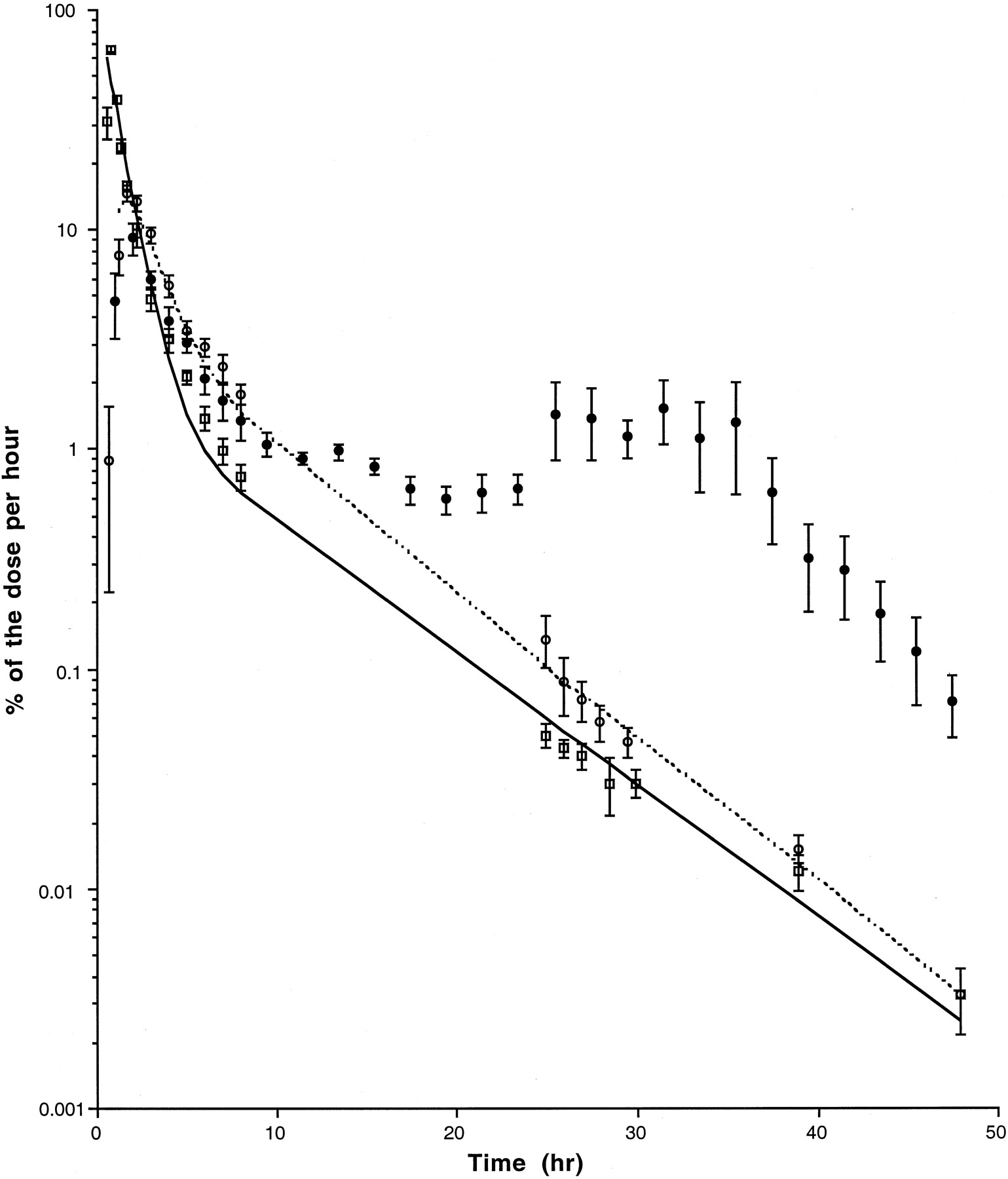
Radioactivity was rapidly and extensively excreted in bile of rats given an i.v. 1 mg/kg dose. Thus, within the first 30 min after administration, 22.4 ± 3.8% of the dose was excreted in bile, and about 80% of the total radioactivity eliminated over 48 h was excreted during the first 8 h. Biliary radioactivity declines were fitted to a two component negative exponential function. The two slopes were calculated to be 1.3/h and 0.15/h, respectively (Fig.2).

Biliary excretion of [14C]1,2-DEB in male rats.
Rates of biliary excretion of radioactivity were calculated at the midpoint of each collection period of bile in rats treated i.v. with 1 mg/kg ([box]; n = 8) or orally with 1 mg/kg dose (○; n = 6) or with 100 mg/kg dose (●;n = 3). The mean equations for individual bile excretion of 1 mg/kg dose administered i.v. or orally were respectively: and
and
The maximum excretion efflux of radioactivity in bile occurred between 1 and 1.5 h after oral administration of 1 and 100 mg/kg, and accounted for 13.0 and 7.4% of the dose per hour, respectively. At the lower dose, the biliary radioactivity decline followed two negative exponential functions. The slope of the second function was not significantly different from that obtained after an i.v. administration.
Although the elimination of radioactivity in bile from rats dosed orally with the highest dose was roughly log-linear up to approximately 8 h, its pattern changed considerably thereafter. Thus, during the 24- to 30-h period collection the quantity of radioactivity excreted per hour was constant, and after that the biliary radioactivity efflux decreased rapidly. This phenomenon was confirmed by a second, independent experiment where the bile of three rats was collected more frequently (Fig. 2).
Radioactivity Excreted by Donor and Recipient Rats.
Rats with crossover bile cannulation were used to determine the extent of the enterohepatic circulation, and to obtain information on the contribution of bile metabolites to the tissue uptake and to the urinary excretion of [14C]1,2-DEB.
Radioactivity excreted within 30 h in the bile of donor rats was calculated to be 63.8% ± 5.1 (n = 5) of the administered dose, which correlated well with the value obtained from single cannulated rats dosed i.v. with 1 mg/kg 1,2-DEB (Table3). Comparison of biliary and fecal data from donors with those of recipient rats gave an estimation of the reabsorption of biliary metabolites. It was found that 94 ± 1% of the biliary metabolites the recipient received were reabsorbed, with only 6% being in the gastrointestinal tract and feces. A large part of the biliary metabolites that were reabsorbed by the receiver rats were then excreted in their own bile.
Excretion and distribution of radioactivity 30 h after [14C]1,2-DEB i.v. administration to male Sprague-Dawley rats with crossover bile cannulation3-a
All tissue concentrations and urinary excretions were higher in donor rats than in recipient rats 30 h after [14C]1,2-DEB dosage. The biliary contribution to both urinary excretion and tissue concentrations was calculated to be about 30% (except for the liver). The results from the experiment directly demonstrated the partial contribution of the biliary metabolites to the brain uptake and urinary excretion of 1,2-DEB.
Analysis of [14C]1,2-DEB Metabolites in Excreta.
Metabolites of 1,2-DEB were analyzed in urine, bile, or fecal homogenates from rats dosed i.v. with 1 mg/kg of [14C]1,2-DEB or dosed orally with 100 mg/kg (Table 4).
Analysis of metabolites in excreta 24 h after administration of [14C]1,2-DEB to male Sprague-Dawley rats4-a
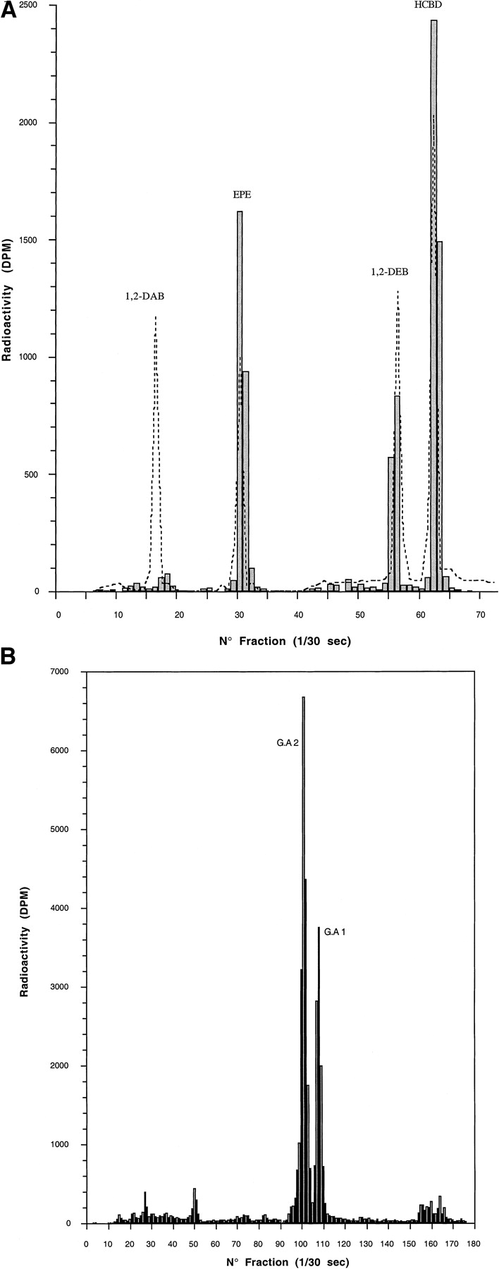
Regardless of the treatment, less than 10% of the radioactivity contained in the urine or bile samples were found as neutral metabolites. One radioactive peak had the same retention time in HPLC as authentic EPE. Neutral metabolites accounted for 25% of the fecal radioactivity. Two major radioactive peaks were eluted at the same retention time as that of 1,2-DEB and EPE standard in feces of rats dosed orally (Fig. 3A). After HPLC-separation, the structure of the radioactive peak that corresponded to EPE was confirmed by GC/MS. Insignificant radioactivity was recovered at a retention time corresponding to a standard of 1,2-DAB.

A, HPLC chromatogram of neutral [14C]1,2-DEB metabolites.
Feces of Sprague-Dawley male rats (n = 5) dosed orally with [14C]1,2-DEB (100 mg/kg) were collected within 24 h. The elution times of the radioactive peaks (▪) of neutral metabolites were compared with those of authentic standards of 1,2-DAB, EPE, and 1,2-DEB analyzed at λ = 263 nm (—). HCBD was used as an internal standard. B, HPLC chromatogram of acidic [14C]1,2-DEB metabolites. Urine of Sprague-Dawley male rats (n = 5) dosed orally with [14C]1,2-DEB (100 mg/kg) were collected within 24 h. GA1 and GA2 accounted for 20 and 37%, respectively, of the urinary radioactivity and have been identified as glucuronide conjugates of EPE.
Approximately 80 to 94% of the urine or bile radioactivity and 10 to 19% of fecal radioactivity were recovered in the fraction corresponding to acidic metabolites. In urine and bile, two major radioactive peaks, GA1 and GA2, were separated by HPLC (Fig. 3B), but were minimal in feces. It is noticeable that the percentages of the metabolites EPE, GA1, and GA2 in urine of non bile duct-cannulated rats dosed i.v. with 1 mg/kg or orally with 100 mg/kg were quite similar. Furthermore, in the urine or bile of these rats, GA1 was lower than GA2.
Enzymatic Hydrolysis of Urine.
A pool of urine collected from rats dosed orally with 100 mg/kg was incubated with β-glucuronidase and analyzed by HPLC. The two urinary metabolites GA1 and GA2 were completely hydrolyzed by a β-glucuronidase from E. coli. After hydrolysis, the radioactivity corresponding to these two metabolites was recovered at the retention time of a standard EPE. In contrast, after hydrolysis of urine by a β-glucuronidase from H. pomatia, only GA1 was readily hydrolyzed (Table 5).
Analysis of urinary metabolites before and after β-glucuronidase hydrolysis
Identification of Glucuronide Conjugates of EPE.
The two isomers separated by HPLC (GA1 and GA2) showed similar mass spectra. The same fragment ions were present; only very minor differences in the relative intensities of some peaks were observed.
The EI spectra were in agreement with the predicted structure but the data were insufficient for identification. The expected molecular ion (M+ 340) was not observed. The main fragment occurred at m/z 133 and was attributed to a 1-(2′-ethylphenyl)ethyl moiety.
The PICI spectra showed peaks at m/z 358 and 323, which were assumed to correspond to the [MNH4]+ and [M+H-H2O]+ ions, respectively.
FAB-MS yielded ions at m/z 363, 358, and 323, attributed to [MNa]+, [MNH4]+, and [M+H-H2O]+. High-resolution FAB-MS of these ions confirmed the glucuronide structure for both isomers.
Discussion
The results of this study indicate that 1,2-DEB is extensively metabolized and rapidly excreted in urine. Thus, about 75% of administered 14C activity was excreted in urine 168 h after a single i.v. administration of a low dose (1 mg/kg) of [14C]1,2-DEB, and 90% of the total radioactivity recovered in urine was excreted within the first 2 days after dosing. However, experiments with rats fitted with a biliary cannula indicated that a large fraction of the [14C]1,2-DEB and/or its metabolites was excreted in bile. The comparison of the biliary excretion of radioactivity over 2 days with that in feces over 7 days suggests that there is an extensive reabsorption of biliary metabolites and an enterohepatic circulation.
Moreover, the experiments involving bile donor and recipient rats indicated that the biliary metabolites underwent several enterohepatic recyclings before being ultimately excreted in urine. Approximately 64% of a 1 mg/kg dose given i.v. was excreted in bile of the donor rats, and then 94% of the biliary metabolites were reabsorbed by the receiver rats. The receiver rats themselves excreted a large part of the absorbed radioactivity in their own bile. The results from the rats linked in a cascade fashion indicated that the first enterohepatic recycling contributed to approximately 30% to the brain uptake and urinary excretion of radioactivity.
The excretion of 1,2-DEB did not seem to be highly dependent on either the route of administration or on the dose. The radioactivity contents in the different excreta from rats dosed i.v. or orally with a 1 mg/kg dose were not significantly different. These results suggested that a low dose of 1,2-DEB was well absorbed from the gastrointestinal tract. In contrast, comparison of the radioactivity in excreta after a single oral administration dose of 1 or 100 mg/kg indicated a possible saturation of the gastrointestinal absorption. This hypothesis is based on the increase in the percentage of the administered dose excreted in feces, with its decrease in urine from rats treated orally at the highest dose. However, the decrease in the gastrointestinal absorption of radioactivity with increasing doses was not confirmed when 1,2-DEB was given repeatedly. Thus, the recovery of the radioactivity expressed as a percentage of the administered dose in the different excreta of rats dosed orally with four daily 100 mg/kg doses was not significantly different from that in excreta of rats dosed orally with 1 mg/kg. In addition, from the radioactivity found in the feces from bile duct-cannulated rats, it was estimated that about 90% of 1 and 100 mg/kg doses given orally were absorbed from the gastrointestinal tract.
If a decrease of gastrointestinal absorption seems unlikely at the highest dose, a comparison of the kinetics of the radioactivity eliminated in bile between the two doses tested suggests a transient saturation of the hepatobiliary excretion at the 100 mg/kg oral dose. The maximum excretion efflux of radioactivity in bile was 2-fold higher at the low dose than that at the highest dose. Additionally, after a single oral or i.v. administration of a 1 mg/kg dose, the biliary excretion declines were fitted to a two component negative exponential functions and showed similar final slopes. In contrast, the flux of the biliary excretion of the radioactivity from rats dosed orally with the highest dose of 1,2-DEB decreased between 1.5 and 16 h, and thereafter slightly increased until 30 h after dosing. As the reabsorption of the biliary metabolites contributed to approximately half of the urinary radioactivity, the lower rate of biliary excretion of the radioactivity might explain that the fraction of the administered dose excreted within the first 24 h decreased as the dose increases (results not shown).
The metabolic profile of 1,2-DEB was qualitatively similar in both the bile and urine of rats given an 1 mg/kg i.v. dose. The fraction of neutral metabolites of 1,2-DEB accounted for less than 10% of the biliary or urinary radioactivity. Insignificant amounts of unchanged 1,2-DEB were recovered in urine or bile, indicating that 1,2-DEB was extensively metabolized. The presence of EPE, a neutral metabolite of 1,2-DEB first identified in urine by Ensminger (1995), was confirmed in this experiment. It also was observed in bile and feces. Yet, this metabolite accounted for less than 2% of the urinary and biliary activity. By contrast, about 80 to 90% of the radioactivity in urine or bile corresponded to polar metabolites of 1,2-DEB. The two main polar metabolites (GA1 and GA2) of 1,2-DEB, which accounted for 57 to 79% of urinary and biliary radioactivity, were assumed to be glucuronide conjugates of EPE. After deglucuronidation of GA1 and GA2 with β-glucuronidase from E. coli, all the radioactivity corresponding to these two metabolites was eluted at the same HPLC retention time as an authentic standard of EPE. Additionally, the EI mass spectra of GA1 and GA2 were similar, and high resolution FAB-MS has confirmed their glucuronide structure.
From the present results, the initial conversion step of the chief metabolic pathway of 1,2-DEB appears to be a hydroxylation of the α-carbon atom of the side chain resulting in EPE, as has been reported for the metabolic conversion of ethylbenzene, which yields 1-phenylethanol, (Mc Mahon and Sullivan, 1966, 1968; Kiese and Lenk, 1974; Sullivan et al., 1976; Engström, 1984; Engström et al., 1984). Optical activity is thus introduced into the metabolism pattern of ethylbenzene by the formation of S(−) andR(+) 1-phenylethanol. This oxidation has been shown to be under stereochemical control, as 1-phenylethanol excreted in rat urine was about a 9:1 mixture of R(+) and S(−) enantiomers (Mc Mahon and Sullivan, 1966). However, previously Smith et al. (1954) claimed that identical amounts of both 1-phenylethanol enantiomers are formed and that they are conjugated in vivo to the same extent. In comparison with ethylbenzene and from the findings that GA1 and GA2 were completely hydrolyzed by β-glucuronidase from E. coli and only GA1 by H. pomatia, it was suggested that GA1 and GA2 were the glucuronide conjugates of the two potential enantiomers of EPE. A high degree of β-glucuronidase stereoselectivity from different sources has been reported previously for diastereoisomeric glucuronides of Oxazepam (Ruelius at al., 1979) and of 3-hydroxy-3 methyloxindole (Smith et al., 1996). The ratio of GA1/GA2, which was about 1:2 in urine of non bile duct-cannulated rats dosed i.v. (1 mg/kg) or orally (100 mg/kg), suggests a stereoselectivity in the metabolism of 1,2-DEB and/or in the biodistribution of its metabolites. At the moment, additional studies are conducted to identify the enantiomeric structure of the two glucuronides and to determine which metabolic step(s) of 1,2-DEB is under stereochemical control. EPE was extensively glucuronoconjugated (GA1 and GA2) and excreted in bile or urine. Reabsorption of the biliary glucuronide conjugates from the gut is more likely to occur after hydrolysis to the lower molecular weight, more lipophilic EPE. Evidence for partial hydrolysis has been obtained by the absence in feces of GA1 and GA2 and the presence of a significant quantity of EPE in the feces of rats dosed i.v.
In a previous report, 1,2-DAB, which has neurotoxic properties, has been identified (but not quantified) by GC/MS in the urine samples of rats given 165 mg/kg 1,2-DEB orally on four consecutive days (Gagnaire et al., 1991). In the present study, insignificant amounts of 1,2-DAB were recovered in urine, bile, or feces of rats dosed with [14C]1,2-DEB. This discrepancy might be a consequence of differences in animal treatment. However, it is known that 1,2-DAB reacts rapidly with amino groups, and it is likely that this metabolite can react in situ with cellular components. Consequently, it can be expected that its urinary excretion level would be rather low. The metabolic scheme for 1,2-DEB in rats shown in Fig.4 is based on the results of Gagnaire et al. (1991), Ensminger et al. (1995), and the present study.
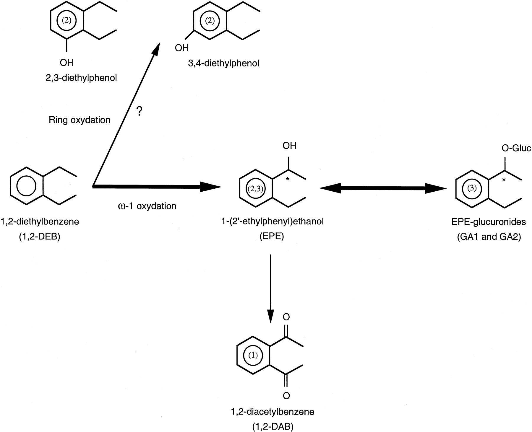
A proposed metabolic scheme of 1,2-DEB in male Sprague-Dawley rats.
Large arrow direction, major metabolic pathway; small arrow direction, minor metabolic. ?, hypothetical pathway; ∗, chiral carbon. 1, fromGagnaire et al., 1991; 2, from Ensminger, 1995; 3, from the present study.
In conclusion, this study has shown that 1,2-DEB is extensively metabolized and mainly excreted in bile or urine as two glucuronide conjugates of EPE. Biliary metabolites were extensively reabsorbed from the gut and ultimately excreted in urine. Except for a transient gastrointestinal absorption and/or hepatobiliary excretion saturation that occurred after an oral administration of a 100 mg/kg dose, no real difference was observed in the excretion or metabolism pattern of [14C]1,2-DEB administered i.v. (1 mg/kg) or orally (1 or 100 mg/kg).
Acknowledgments
We thank F. Canel, P. Chaumont, and M.C. Grandclaude for their technical assistance and M. Roussel and C. Caël for their expert secretarial services.
Footnotes
-
Send reprint requests to: Dr. Jean-Paul Payan, Institut National de Recherche et de Sécurité, Avenue de Bourgogne, B.P. No. 27, 54501 Vandoeuvre Cedex, France. E-mail: payanjp{at}inrs.fr
- Abbreviations used are::
- DEB
- diethylbenzene
- 1,2-DAB
- 1,2-diacetylbenzene
- 1,2-DEB
- 1,2-diethylbenzene
- [14C]1,2-DEB
- 1,2-diethyl[U-14C]benzene
- EI
- electron impact
- EPE
- 1-(2′-ethylphenyl)ethanol
- Et2O
- diethyl ether
- FAB
- fast atom bombardment
- GA1 and GA2
- glucuronide conjugates of EPE
- GC/MS
- gas chromatography/mass spectrometry
- MS
- mass spectrometry
- [14C]HCBD
- [14C]hexachlorobutadiene
- PICI
- positive ion chemical ionization
- Received May 10, 1999.
- Accepted August 11, 1999.
- The American Society for Pharmacology and Experimental Therapeutics


