


Abstract
The present study was designed to assess both preventive and therapeutic effects of (S)-1-(2-Hydroxyethyl)-4-methyl-N-[4-(methylsulfonyl) phenyl]-5-[2-(trifluoromethyl) phenyl]-1H-pyrrole-3-carboxamide (CS-3150), a novel nonsteroidal mineralocorticoid receptor antagonist, on renal injury in deoxycorticosterone acetate (DOCA)/salt-induced hypertensive rats (DOCA rats). From 7 weeks of age, DOCA was subcutaneously administered once a week for 4 weeks to uninephrectomized rats fed a high-salt diet. In experiment 1, CS-3150 (0.3–3 mg/kg) was orally administered once a day for 4 weeks coincident with DOCA administration. In experiment 2, after establishment of renal injury by 4 weeks of DOCA/salt loading, CS-3150 (3 mg/kg) was orally administered once a day for 4 weeks with or without continuous DOCA administration. In experiment 1, DOCA/salt loading significantly increased systolic blood pressure (SBP), which was prevented by CS-3150 in a dose-dependent manner. Development of renal injury (proteinuria, renal hypertrophy, and histopathological changes in glomeruli and tubule) was also suppressed by CS-3150 with inhibition of mRNA expression of fibrosis, inflammation, and oxidative stress markers. In experiment 2, under continuous DOCA treatment, CS-3150 clearly ameliorated existing renal injury without lowering SBP, indicating that CS-3150 regressed renal injury independent of its antihypertensive action. Moreover, CS-3150 treatment in combination with withdrawal of DOCA showed further therapeutic effect on renal injury accompanied by reduction in SBP. These results demonstrate that CS-3150 not only prevents but also ameliorates hypertension and renal injury in DOCA rats. Therefore, CS-3150 could be a promising agent for the treatment of hypertension and renal disorders, and may have potential to promote regression of renal injury.
Introduction
Mineralocorticoid receptor (MR) was originally recognized to be expressed in epithelial cells of the distal tubule and collecting duct in the kidney, and to play a role in the regulation of electrolyte homeostasis, body fluid, and blood pressure (Funder, 1995). In addition to such classic actions, it has been reported that MR is also expressed in other cells in the kidney, such as podocytes (Shibata et al., 2007), mesangial cells (Nishiyama et al., 2005), and fibroblasts (Nagai et al., 2005), and MR activation by endogenous ligands (e.g., aldosterone) is involved in the pathogenesis of renal disorder via direct enhancement of fibrosis (Brem et al., 2011), inflammation (Siragy and Xue, 2008), and oxidative stress (Patni et al., 2007). In fact, numerous clinical studies using spironolactone and eplerenone, which are clinically available MR antagonists, have demonstrated that MR blockade is an effective strategy for treatment of chronic kidney disease (Sato et al., 2005; Mehdi et al., 2009; Boesby et al., 2011) as well as hypertension (Chapman et al., 2007; Calhoun and White, 2008).
Recently, we discovered (S)-1-(2-hydroxyethyl)-4-methyl-N-[4-(methylsulfonyl) phenyl]-5-[2-(trifluoromethyl) phenyl]-1H-pyrrole-3-carboxamide (CS-3150), a novel nonsteroidal MR antagonist. CS-3150 is a selective and highly potent MR antagonist with long-lasting MR antagonism after oral administration (Arai et al., 2015). It is well known that deoxycorticosterone acetate (DOCA), a synthetic mineralocorticoid, induces hypertension and renal injury in combination with salt loading to rats (DOCA rats), and this hypertensive rat is widely used as an MR-mediated disease model to evaluate pharmacological effects of MR antagonists or other drugs (Klanke et al., 2008; Seifi et al., 2010). We have previously reported that CS-3150 inhibits elevation in systolic blood pressure (SBP) in DOCA rats (Arai et al., 2015); however, we have not examined the effects of CS-3150 on renal injury. In addition, it remains unclear whether CS-3150 elicits therapeutic efficacy in the established hypertension and renal injury in DOCA rats. Therefore, in the present study, two independent experiments were conducted to investigate the preventive and therapeutic effects of CS-3150 on hypertension and renal injury in DOCA rats.
Materials and Methods
Test Compounds.
CS-3150 was synthesized at Medicinal Chemistry Research Laboratories, Daiichi Sankyo Co., Ltd. (Tokyo, Japan), as described in patent no. WO2008126831. The chemical structure of CS-3150 is shown in Fig. 1.

Chemical structure of CS-3150.
Experimental Protocol.
All procedures involving animal use in these experiments were carried out in accordance with the guidelines of the Institutional Animal Care and Use Committee of Daiichi Sankyo Co., Ltd. Five-week-old male Wistar Kyoto (WKY/Izm) rats were purchased from Funabashi Farm Co., Ltd. (Chiba, Japan). The rats were maintained in a room at 50 ± 10% relative humidity and 23 ± 2°C under a 12-hour light/dark cycle, and were allowed free access to food (FR-2; Funabashi Farm Co., Ltd.) and water.
In experiment 1 (preventive study), 6-week-old male WKY/Izm rats were anesthetized with isoflurane inhalation, and the left kidney was removed. After 1-week recovery (at 7 weeks of age), the rats were divided into five groups: control, vehicle, and three groups of CS-3150 treatments. A 20-mg/kg dose of DOCA, suspended in 0.5% carboxymethylcellulose (CMC; Wako Pure Chemical Industries, Ltd., Osaka, Japan), was subcutaneously administered once a week for 4 weeks. In control rats, 0.5% CMC was administered instead of DOCA. The rats were fed a high-salt diet (FR-2 containing 4% NaCl; Funabashi Farm Co., Ltd.) during the study. To assess the preventive effect of CS-3150, vehicle (0.5% methylcellulose [MC]; Wako Pure Chemical Industries, Ltd.) or CS-3150 (0.3, 1, and 3 mg/kg) was orally administered once a day for 4 weeks.
In experiment 2 (therapeutic study), hypertension and renal damage were established by 4-week DOCA/salt loading as conducted in experiment 1. At 11 weeks of age, the control rats (0.5% CMC–treated rats) were divided into two subgroups, and the DOCA-treated rats were divided into five subgroups. One of each subgroup was dissected according to the following procedure as the baseline. Two subgroups of DOCA-treated rats received a further 4-week administration of DOCA, whereas the other two received 0.5% CMC instead. To assess the therapeutic effect of CS-3150, vehicle (0.5% MC) or CS-3150 (3 mg/kg) was orally administered once a day for 4 weeks. Regarding a subgroup of control rats, 0.5% CMC was received once a week, and vehicle (0.5% MC) was orally administered once a day for a further 4 weeks. All rats were fed a high-salt diet (FR-2 containing 4% NaCl; Funabashi Farm Co., Ltd.) during the study.
Measurement of Blood Pressure and Urinary Parameters.
SBP was measured by the tail-cuff method (BP-98A; Softron Co., Ltd., Tokyo, Japan) in awake, restrained animals at 7, 9, and 11 weeks of age in experiment 1, and at 7, 11, 13, and 15 weeks of age in experiment 2.
Rats were individually placed in a metabolic cage, and urine was collected for 24 hours at 7 and 11 weeks of age in experiment 1, and at 7, 11, 13, and 15 weeks of age in experiment 2. Urinary volume (UV) was measured, and the urine samples were centrifuged (1820 × g for 15 minutes). The supernatant was used for measurement of protein concentration by an automated clinical analyzer (BiOLiS 24i Premium; Tokyo Boeki Machinery Ltd., Tokyo, Japan), and urinary protein (UP) excretion for 24 hours was calculated. Urinary sodium (Na+) and potassium (K+) concentrations were also measured using an electrolyte analyzer (STAX-2; Techno Medica Co., Ltd., Kanagawa, Japan), and both excretions for 24 hours were calculated (Supplemental Figs. 1 and 2). In addition, urinary MCP-1 concentration was measured by an enzyme-linked immunosorbent assay kit (R&D Systems, Minneapolis, MN), and its excretion for 24 hours was calculated (Supplemental Tables 1 and 2).
Necropsy.
In experiment 1, at 11 weeks of age, each rat was anesthetized by isoflurane inhalation, and after blood sampling from the abdominal aorta, the right kidney was excised and weighed. A part of the right kidney was placed in RNA stabilization reagent (RNAlater; Invitrogen, Carlsbad, CA) and stored at −80°C until total RNA isolation. The rest of the right kidney was sectioned into 2- to 3-mm slices and fixed in 10% neutral buffered formaldehyde solution (Wako Pure Chemical Industries, Ltd.). In experiment 2, necropsy was performed at 11 weeks of age in some of the rats as the baseline and at 15 weeks of age on the remaining rats, as described earlier.
Relative mRNA Expression Analysis by Real-Time Polymerase Chain Reaction.
Relative gene expression in the kidney was determined by quantitative real-time polymerase chain reaction (PCR) as follows. Total RNA was extracted using TRIzol Reagent (Invitrogen), and the cDNA was synthesized by a First-Strand cDNA Synthesis Kit (GE Healthcare, Buckinghamshire, UK) with random hexamer primers. Quantitative PCR was performed using the cDNA by TaqMan Gene Expression Assays (Applied Biosystems, Foster City, CA) and TaqMan universal PCR master mix according to the manufacturer’s instructions. The following assays were used: TGF-β1 (Rn00572010_m1), collagen 1a1 (Col1a1; Rn01463848_m1), interleukin-6 (IL-6; Rn01410330_m1), MCP-1 (Rn00580555_m1), p47phox (Rn00586945_m1), p67phox (Rn01759079_m1), and peptidylprolyl isomerase B (Rn03302274_m1). PCR reactions were performed using a 7900 HT Fast Real Time PCR system (Applied Biosystems). The data were analyzed automatically using SDS 2.4 software (Applied Biosystems) to determine the expression levels. Relative mRNA levels of each gene were calculated by normalizing them to the peptidylprolyl isomerase B mRNA level.
Histopathological Examination of Kidney.
The excised right kidney fixed in 10% neutral buffered-formaldehyde solution was embedded in paraffin. Sections were stained with periodic acid methenamine silver for the following histopathological examination. Severity of glomerulosclerosis was semiquantitatively evaluated in 30 glomeruli in each specimen according to the criteria developed by Uehara et al. (1993). The extent of glomerulosclerosis was graded from 0 to 4 as follows: 0, normal; 1, <25% of sclerosis; 2, 25–50% of sclerosis; 3, 50–75% of sclerosis; and 4, >75% of sclerosis. Severity of tubular injury of both the cortex and medulla was also semiquantitatively evaluated in 10 randomly selected microscope fields (×200) in each specimen according to the criteria developed by Uehara et al. (1993). The extent of tubular injury was graded from 0 to 4 as follows: 0, no lesion; 1, very mild focal dilatation of tubules; 2, large number of dilated tubules with widening of interstitium; 3, fairly extensive dilatation of tubules with cystic formation and widening of interstitium; and 4, complete atrophy of tubules.
Statistical Analysis.
Data are expressed as the mean ± S.E.M. The comparison between two groups was evaluated by Student’s t test or Aspin-Welch’s t test. For multiple comparisons, Dunnett’s test was performed. A P value of less than 0.05 was considered to be statistically significant. All calculations were conducted using SAS System Release 8.2 and 9.2 (SAS Institute Inc., Tokyo, Japan).
Results
Experiment 1
SBP and UP Excretion.
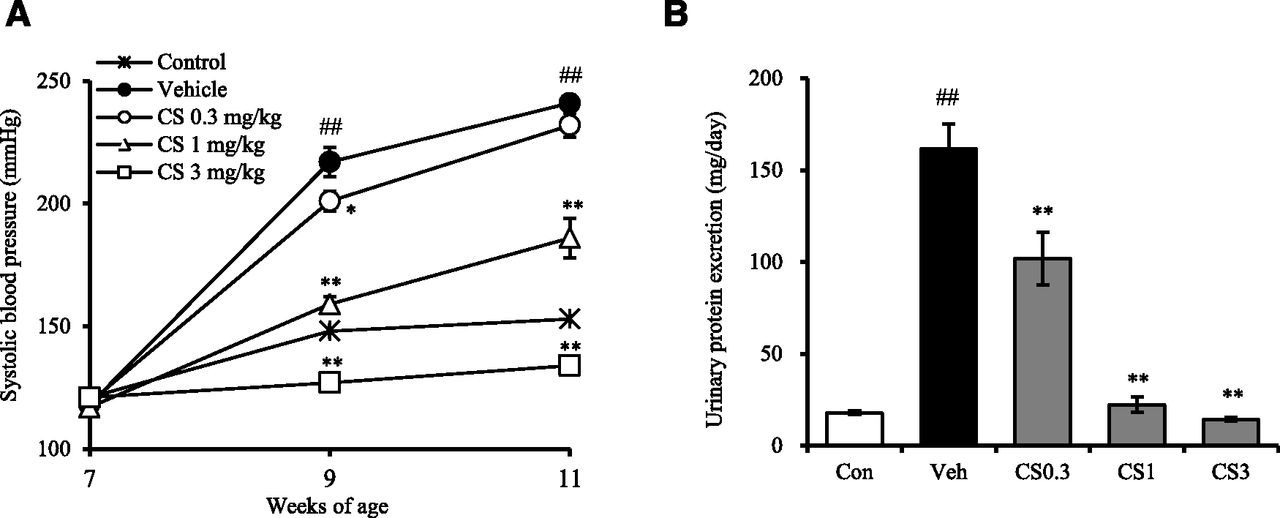
DOCA/salt loading to WKY/Izm rats induced progressive elevation of SBP (Fig. 2A). Treatment with CS-3150 suppressed SBP elevation in a dose-dependent manner, and the antihypertensive effect at 11 weeks of age was significant at 1 and 3 mg/kg (Fig. 2A). UP excretion was also increased in the vehicle group, and was prevented by CS-3150 (Fig. 2B). Interestingly, 1 mg/kg CS-3150 completely blocked the increase in UP excretion while only partially suppressing SBP elevation (Fig. 2, A and B).

Effects of CS-3150 (CS) on systolic blood pressure and urinary protein excretion in DOCA/salt-induced hypertensive rats (in experiment 1). From 7 weeks of age, DOCA was subcutaneously administered once a week for 4 weeks to uninephrectomized rats fed a high-salt (4% NaCl) diet. CS-3150 (0.3–3 mg/kg) was orally administered once a day for 4 weeks from the start date of DOCA administration. (A) Systolic blood pressure was measured at 7, 9, and 11 weeks of age. (B) Urine was collected for 24 hours at 11 weeks of age, and urinary volume and protein concentration were measured. Urinary protein excretion for 24 hours was calculated. Con, control group (no DOCA administered); CS0.3, 0.3 mg/kg CS-3150–treated group; CS1, 1 mg/kg CS-3150–treated group; CS3, 3 mg/kg CS-3150–treated group; Veh, vehicle-treated group. Data are expressed as the mean ± S.E.M. (N = 6 in each group). ##P < 0.01 versus control; *P < 0.05 versus vehicle; **P < 0.01 versus vehicle.
Other Urinary Parameters.
UV was increased in the vehicle group, and was prevented by CS-3150 (Supplemental Fig. 1A). Urinary Na+ and K+ excretions were also increased in the vehicle group; however, there were no significant differences between the vehicle- and CS-3150–treated groups (Supplemental Fig. 1, B and C).
Urinary MCP-1 excretion was increased in the vehicle, and was prevented by CS-3150 (Supplemental Table 1).
Kidney Weight and Histology.
Kidney weight was markedly increased in the vehicle group (Fig. 3A). In addition, DOCA/salt loading to rats resulted in marked glomerular and tubular injury, such as glomerulosclerosis, dilation of tubules, widening of interstitium, and cystic formation (Figs. 3, B–D, and 4). CS-3150 dose-dependently prevented both renal hypertrophy and histopathological changes.

Effects of CS-3150 on kidney weight and histopathological changes in the kidney of DOCA/salt-induced hypertensive rats (in experiment 1). From 7 weeks of age, DOCA was subcutaneously administered once a week for 4 weeks to uninephrectomized rats fed a high-salt (4% NaCl) diet. CS-3150 (0.3–3 mg/kg) was orally administered once a day for 4 weeks from the start date of DOCA administration. (A) At 11 weeks of age, the right kidney was removed under anesthesia and weighed. The kidney weight/body weight (BW) ratio was calculated. (B–D) The sections of excised right kidney were stained with periodic acid methenamine silver, and severity of glomerulosclerosis and tubular injury (cortex and medulla) was semiquantitatively evaluated. Con, control group (no DOCA administered);CS0.3, 0.3 mg/kg CS-3150–treated group; CS1, 1 mg/kg CS-3150–treated group; CS3, 3 mg/kg CS-3150–treated group; Veh, vehicle-treated group. Data are expressed as the mean ± S.E.M. (N = 6 in each group). ##P < 0.01 versus control; **P < 0.01 versus vehicle.

Representative photomicrographs showing glomerulosclerosis and tubular injury in DOCA/salt-induced hypertensive rats (in experiment 1). CS, CS-3150.
mRNA Expression in the Kidney.
In the kidney of DOCA rats, mRNA expression levels of profibrotic markers, such as TGF-β1 and Col1a1, and proinflammatory cytokines, such as IL-6 and MCP-1, were markedly increased compared with the control rats (Table 1). NADPH oxidase is known to cause reactive oxygen species production, and the mRNA expression levels of NADPH oxidase subunits, such as p47phox and p67phox, were also increased in the kidney of DOCA rats (Table 1). Treatment with CS-3150 clearly inhibited the induction of mRNA expression of these markers in a dose-dependent manner.
Effects of CS-3150 on mRNA expression levels in the kidney in DOCA/salt-induced hypertensive rats (in experiment 1)
Data are relative mRNA levels of each gene calculated by normalizing them to the peptidylprolyl isomerase B mRNA level, and are expressed as the mean ± S.E.M. (N = 6 in each group).
Experiment 2
SBP and UP Excretion.
Consistent with experiment 1, SBP in the vehicle group was significantly higher than that in the control group at 11 weeks of age, and additional 4-week administration of DOCA induced further SBP elevation (Fig. 5A). Under continuous DOCA treatment, CS-3150 inhibited further SBP elevation at 13 weeks of age, but SBP at 15 weeks of age was equivalent to that in the vehicle group (Fig. 5A). Withdrawal of DOCA treatment did not show any significant difference in SBPs at either 13 or 15 weeks of age. CS-3150 treatment without DOCA administration dramatically lowered SBP to the same level as the control group.

Effects of CS-3150 on systolic blood pressure and urinary protein excretion in DOCA/salt-induced hypertensive rats (in experiment 2). From 7 weeks of age, DOCA was subcutaneously administered once a week for 4 weeks to uninephrectomized rats fed a high-salt (4% NaCl) diet. From 11 weeks of age, CS-3150 (CS; 3 mg/kg) was orally administered once a day for 4 weeks with or without continuous DOCA administration. (A) Systolic blood pressure was measured at 7, 11, 13, and 15 weeks of age. (B) Urine was collected for 24 hours at 7, 11, 13, and 15 weeks of age, and urinary volume and protein concentration were measured. Urinary protein excretion for 24 hours was calculated. Data are expressed as the mean ± S.E.M. (N = 6 in each group). ##P < 0.01 versus control; * P < 0.05 versus vehicle; **P < 0.01 versus vehicle.
UP excretion was markedly increased at 11 weeks of age, and additional 4-week administration of DOCA caused a further increase in UP excretion (Fig. 5B). CS-3150 significantly reduced UP excretion under continuous DOCA treatment (Fig. 5B). Withdrawal of DOCA treatment gradually decreased UP excretion, and CS-3150 treatment without DOCA administration elicited a further reduction in UP excretion (Fig. 5B).
Other Urinary Parameters.
UV was increased in the vehicle group, and was gradually decreased by CS-3150 treatment and/or withdrawal of DOCA treatment (Supplemental Fig. 2A). CS-3150 treatment and/or withdrawal of DOCA treatment did not show any significant effect on urinary Na+ and K+ excretions (Supplemental Fig. 2, B and C).
At 15 weeks of age, urinary MCP-1 excretion was reduced by CS-3150 treatment and/or withdrawal of DOCA treatment, compared with the vehicle group (Supplemental Table 2).
Kidney Weight and Histology.
In the vehicle group, kidney weight was significantly increased compared with the control group at 11 weeks of age, and DOCA treatment for a further 4 weeks maintained this renal hypertrophy until 15 weeks of age (Fig. 6A). Withdrawal of DOCA treatment reduced kidney weight to the control level, meaning that MR activation contributes to renal hypertrophy in this model. In fact, treatment with CS-3150 markedly reduced kidney weight regardless of DOCA treatment.

Effects of CS-3150 (CS) on kidney weight and histopathological changes in the kidney of DOCA/salt-induced hypertensive rats (in experiment 2). From 7 weeks of age, DOCA was subcutaneously administered once a week for 4 weeks to uninephrectomized rats fed a high-salt (4% NaCl) diet. From 11 weeks of age, CS-3150 (3 mg/kg) was orally administered once a day for 4 weeks with or without continuous DOCA administration. (A) At 11 or 15 weeks of age, the right kidney was removed under anesthesia and weighed. The kidney weight/body weight (BW) ratio was calculated. (B–D) The sections of excised right kidney were stained with periodic acid methenamine silver, and severity of glomerulosclerosis and tubular injury (cortex and medulla) was semiquantitatively evaluated. Data are expressed as the mean ± S.E.M. (N = 6 in each group). $$P < 0.01 versus control at 11 weeks of age; ##P < 0.01 versus control at 15 weeks of age; bbP < 0.01 versus vehicle at 11 weeks of age; **P < 0.01 versus vehicle at 15 weeks of age.
Consistent with experiment 1, marked histopathological changes in kidney were observed in the vehicle group at 11 weeks of age, and further treatment with DOCA for 4 weeks exacerbated these changes, especially glomerulosclerosis (Figs. 6, B–D, and 7). Interestingly, under continuous treatment of DOCA, CS-3150 not only inhibited these changes but also clearly ameliorated them, indicating that CS-3150 induced regression of renal injury. Withdrawal of DOCA treatment also resulted in a similar amelioration, and CS-3150 treatment without DOCA administration elicited further improvement of renal histology.

Representative photomicrographs showing glomerulosclerosis and tubular injury in DOCA/salt-induced hypertensive rats (in experiment 2). CS, CS-3150.
mRNA Expression in the Kidney.
Consistent with experiment 1, mRNA expression levels of profibrotic markers (TGF-β1 and Col1a1), proinflammatory cytokines (IL-6 and MCP-1), and NADPH oxidase subunits (p47phox and p67phox) were markedly increased in the kidney of DOCA rats compared with the control rats at 11 weeks of age, and the same increase was noted at 15 weeks of age (Table 2). Under continuous DOCA treatment, CS-3150 clearly reduced the mRNA expression of some of these markers. Withdrawal of DOCA treatment also showed a similar effect, and CS-3150 treatment without DOCA administration elicited a further decrease in mRNA expression.
Effects of CS-3150 on mRNA expression levels in the kidney in DOCA/salt-induced hypertensive rats (in experiment 2)
Data are relative mRNA levels of each gene calculated by normalizing them to the peptidylprolyl isomerase B mRNA level, and are expressed as the mean ± S.E.M. (N = 6 in each group).
Discussion
This is the first report that shows a therapeutic effect of CS-3150 as well as a preventive effect on hypertension and renal injury in DOCA/salt-hypertensive rats. CS-3150 (0.3–3 mg/kg) dose-dependently prevented the elevation of SBP induced by DOCA/salt loading. CS-3150 also suppressed the development of renal injury (i.e., proteinuria, renal hypertrophy, and histopathological changes in glomeruli and tubule) with inhibition of gene expression related to fibrosis, inflammation, and oxidative stress. In addition, CS-3150 at 3 mg/kg clearly ameliorated existing renal injury without affecting SBP, even under continuous DOCA treatment, indicating that CS-3150 has the potential to regress established renal injury in DOCA rats. Moreover, CS-3150 in combination with withdrawal of DOCA treatment showed further therapeutic effect on renal injury with reduction in SBP. Therefore, these are the first results to demonstrate that CS-3150 elicited regression of overt proteinuria with histologic restoration of glomerular and tubular damage in DOCA rats.
In this study, repeated administration of CS-3150 for 4 weeks significantly inhibited the onset of hypertension in the DOCA rats, which was consistent with the results of our previous study for 2 weeks (Arai et al., 2015). SBP was measured by the tail-cuff method, which is a little less accurate than the telemetry method. Nevertheless, this would not be a major issue since the dose-dependent antihypertensive effect of CS-3150 was clearly observed. DOCA/salt loading also caused marked proteinuria, renal hypertrophy, and glomerular and tubular injury, which were significantly prevented by treatment with CS-3150. Interestingly, significant suppression of renal injury in the absence of a blood pressure–lowering effect was detected at 0.3 mg/kg CS-3150, suggesting that the renal protective effect of CS-3150 could not be explained simply by its antihypertensive action. Accumulating evidence has shown that aldosterone/MR signaling directly contributes to the pathogenesis of renal damage such as tubulointerstitial fibrosis through collagen accumulation (Sun et al., 2006; Diah et al., 2008), increased expression of proinflammatory cytokines in the kidney (Blasi et al., 2003; Ikeda et al., 2009), and renal reactive oxygen species generation by an NADPH oxidase–dependent mechanism (Beswick et al., 2001; Nishiyama et al., 2004). In the current study, upregulation of mRNA for several markers related to fibrosis (TGF-β1 and Col1a1), inflammation (MCP-1 and IL-6), and oxidative stress (p47phox and p67phox) was observed in the kidney of DOCA rats, and CS-3150 treatment inhibited these changes. We also confirmed that increase in urinary MCP-1 excretion was inhibited by CS-3150 treatment (Supplemental Table 1). Taken together, the preventive effect of CS-3150 on renal injury could be due to not only antihypertensive but also direct renal protective effects through antifibrotic, anti-inflammatory, and antioxidative actions.
So far, renal injury such as glomerulosclerosis and tubular injury has been considered to be irreversible once established (Klahr, 1999; Phillips et al., 1999). However, recent clinical and nonclinical studies have shown that it can be reversed (Adamczak et al., 2003; Gaede et al., 2004; Rossing et al., 2005; Teles et al., 2009). In fact, regression of renal injury has been reported to be achieved by intensive intervention against the causes of renal injury, such as hypertension, diabetes, and hyperlipidemia (Hovind et al., 2001; Fioretto et al., 2006; Zoja et al., 2010). Since renal injury in DOCA rats is caused mainly by MR activation, CS-3150 treatment in DOCA rats with established renal injury could mimic the intensive intervention described earlier. Surprisingly, CS-3150 elicited a marked reduction in proteinuria and kidney weight, and dramatically reversed glomerulosclerosis and tubular injury with only a slight effect on blood pressure, indicating that CS-3150 treatment restored the established renal damage by DOCA/salt loading independent of its antihypertensive action. There is some speculation about the mechanisms for this reversal of renal injury. It has been reported that degradation of extracellular matrix protein would be directly involved in the regression of sclerosis lesion in the kidney by an angiotensin-converting enzyme inhibitor (Remuzzi et al., 2006) or angiotensin receptor blocker (Boffa et al., 2003). In these reports, a decrease in expression of plasminogen activator inhibitor-1 (PAI-1), which is known as an inhibitor of matrix degradation (Ma et al., 2000), was observed in the kidney, resulting in increased metalloproteinase activity and decreased expression of TGF-β1 and collagen types I and IV. In the present study, increased mRNA expression of TGF-β1 and Col1a1 in the kidney was clearly downregulated by CS-3150 treatment. Thus, in the kidney of DOCA rats, degradation of once accumulated extracellular matrix protein may occur, at least in part, through inhibition of PAI-1 by CS-3150 treatment. Brown et al. (2000) described the involvement of PAI-1 in aldosterone-induced renal injury, which could also support this hypothesis. Several reports have recently suggested that an injured kidney attempts to repair and regenerate in both its glomerular and tubular compartments, and stem/progenitor cells could contribute to this process through migration of adjacent undamaged cells (Choi et al., 2009; Benigni et al., 2010; Romagnani et al., 2013). For example, it has been shown that progenitor-like cells are localized in the proximal and distal tubule and migrate to the damaged lesion following ischemia (Maeshima et al., 2003). It has also been reported that transplantation of bone marrow cells improves renal function by replacement of defective podocytes in mice with Alport syndrome (Prodromidi et al., 2006). Although the precise involvement of these cells remains unclear in the present study, CS-3150 may enhance the renal tissue repair process through direct abolishing of renal MR activation by an exogenous DOCA. Specific experiments would be necessary to clarify this speculation.
Additionally, CS-3150 treatment in combination with DOCA withdrawal resulted in further regression of renal injury accompanied by SBP reduction. Besides the classic role of MR in the kidney for blood pressure control, it has been shown that extrarenal MR (e.g., brain and vasculature) is involved in the regulation of blood pressure (Oki et al., 2012; Barrett et al., 2013). For example, Rahmouni et al. (2002) reported that intracerebroventricular administration of an MR antagonist decreased SBP in DOCA rats with developed hypertension. Experimental studies using transgenic mouse models, such as smooth muscle cell–specific MR knockout mice (McCurley et al., 2012) and endothelial MR–overexpressing mice (Rickard et al., 2014), have supported the direct involvement of vascular MR in blood pressure control. From these findings, we speculate that not only renal but also extrarenal MR activation in DOCA rats could be completely inhibited by the combination of CS-3150 treatment and withdrawal of DOCA, resulting in improvement of both renal injury and hypertension to the same level as control rats. Additional experiments would be needed to test this hypothesis.
Induction of urinary Na+ excretion (i.e., diuretic action) is known as a mechanism-based action of MR antagonists (Muller et al., 2003). However, several reports have suggested that it is not easy to assess the diuretic action of MR antagonists, especially in high-salt-induced hypertension model rats (Chabert et al., 1984; Jiménez et al., 1988). Indeed, an increase in urinary Na+ excretion was not noted in CS-3150–treated DOCA rats in the current study, although the precise cause remains unclear (Supplemental Figs. 1 and 2). Also, the result of UV was well correlated with those of SBP and UP excretion, and we think that this simply reflects the inhibitory effects of CS-3150 on DOCA/salt-induced pathology.
Recently, Rafiq et al. (2014) demonstrated that high-salt loading to Dahl salt-sensitive hypertensive rats induced renal injury that was reversed by switching to a normal salt diet (Rafiq et al., 2014). In another report, it has been shown that, in an adenine-loaded rat, which is widely used as a model for renal failure, cessation of the adenine diet led to the reversal of renal damage (Shuvy et al., 2011). In the present study, in DOCA rats with established renal injury, withdrawal of DOCA administration itself reversed renal injury. These observations suggest that elimination of a major contributor to the pathology (i.e., excess salt intake, adenine diet, or DOCA treatment) could lead to regression of renal injury. Although multiple causes are intricately involved in the pathogenesis of renal injury in a clinical setting (Macisaac et al., 2014; Drawz and Rahman, 2015), these findings support the concept that identification of the main pathways involved in kidney injury and targeted intensive treatment may lead to regression of renal injury.
In conclusion, CS-3150 not only prevented the onset of hypertension and renal injury, but also reversed them following their establishment in DOCA rats. In particular, to the best of our knowledge, this is the first investigation on the regression of renal injury in DOCA rats, and CS-3150 clearly exerted a regressive effect on overt proteinuria accompanied by histologic restoration of glomerular and tubular damage. Therefore, CS-3150 could be a promising agent for the treatment of hypertension and renal disorders, and may have potential as a therapeutic approach to renal injury.
Acknowledgments
The authors thank Shin Nippon Biomedical Laboratories, Ltd., Drug Safety Research Laboratories (Kagoshima, Japan) for their expert experiments.
Authorship Contributions
Participated in research design: Arai, Morikawa, Ubukata, Homma.
Conducted experiments: Arai, Morikawa, Ubukata, Homma.
Contributed new reagents or analytic tools: Tsuruoka.
Performed data analysis: Arai, Morikawa, Ubukata, Homma.
Wrote or contributed to the writing of the manuscript: Arai, Homma.
Footnotes
- Received May 5, 2016.
- Accepted June 30, 2016.
↵
 This article has supplemental material available at jpet.aspetjournals.org.

Abbreviations
- CMC
- carboxymethylcellulose
- Col1a1
- collagen 1a1
- CS-3150
- (S)-1-(2-hydroxyethyl)-4-methyl-N-[4-(methylsulfonyl) phenyl]-5-[2-(trifluoromethyl) phenyl]-1H-pyrrole-3-carboxamide
- DOCA
- deoxycorticosterone acetate
- IL-6
- interleukin-6
- MC
- methylcellulose
- MR
- mineralocorticoid receptor
- PAI-1
- plasminogen activator inhibitor-1
- SBP
- systolic blood pressure
- UP
- urinary protein
- UV
- urinary volume
- WKY/Izm
- Wistar Kyoto
- Copyright © 2016 by The American Society for Pharmacology and Experimental Therapeutics


